Picture risks for breast cancer

I’ve been diagnosing breast cancer for 35 years – this is what causes real risk

By Rosie Fitzmaurice Freelance journalist specialising in health.


Zoe Winters, a consultant oncoplastic breast surgeon and specialist, says that breast cancer screening should be more personalised

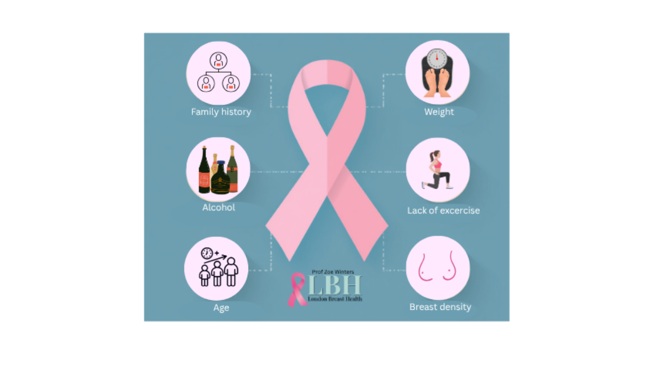
Breast cancer is the most commonly diagnosed cancer among women worldwide. One in seven women in the UK will develop it in their lifetime. Breast cancer risk is complex and influenced by a combination of factors, including genetics, lifestyle and environment.

Not all breast cancers are preventable, but breast specialist Zoe Winters, a consultant oncoplastic breast cancer surgeon at London Breast Health and honorary associate professor at University College London, says one of the most important ways a woman can protect herself against the disease is to understand her baseline risk, to attend screenings and be aware of lifestyle factors which can influence risk.

Here, she shares the things she wants every woman to know.

Your risk increases with age

Age is the single biggest independent risk factor for developing breast cancer. “From age 50, breast cancer risks increase exponentially – the majority (about 80 per cent) occur from this age – and that continues on a steep curve until about 65, when it plateaus,” Winters explains. “Though older women are still at the very highest risk.”

For context, around one in five cancers occur in women under 50 (which works out to be about 10,000 women per year in the UK). While about four per cent of breast cancers occur in women under 40. “Young onset breast cancer, in women [under] 45 tends to be characteristically more biologically aggressive and is typically diagnosed later,” she says.

Family history is important

“In clinic, after age, my next question is ‘Have you ever had breast cancer before?’ Because this automatically puts you into a ‘high risk’ group,” Winters continues. “Family history is very important, too.” This means how many first-degree relatives (parents, siblings) and second-degree (aunts, uncles or grandparents) have had breast and/or ovarian cancer, as well as pancreatic and prostate cancers, on both the maternal and paternal sides.

“Most women (90 per cent) who get breast cancer have not inherited gene mutations, such as BRCA1 and BRCA2. However, it is thought that around five to 10 per cent of women have a specific gene mutation, and 30 per cent of these have no family history.”

Hormonal factors can influence risk

Hormones, such as oestrogen and progesterone, can influence your risk. There has been long-held confusion around whether or not taking HRT raises your risk. The NHS states that taking combined HRT (oestrogen and progesterone) can slightly increase risk, but the increase is small, and for many the benefits outweigh the risks. Meanwhile, there is little or no increase in risk from taking oestrogen-only HRT.

“Modern forms of HRT can offer health benefits, providing protection against cardiovascular disease and osteoporosis, for example,” Winters says. “Ideally, I’d advise taking bio-identical transdermal oestrogen and using a preferred low dose, and/or cyclical micronised progesterone or intra-uterine progesterone like a Mirena coil. Ultimately, it’s important to take into account your individual risk factors if taking HRT for more than five years.”

Taking oral contraception may also slightly increase your risk of breast cancer but the risk reduces once you stop taking it. Other factors, such as not having children, having your first full-term pregnancy after the age of 30 or not breastfeeding, can also raise your lifetime risk.

This is because pregnancy and breastfeeding pause the menstrual cycle, or the cycling of estrogen and progesterone, which stimulates the breast duct cells, therefore reducing the amount of hormones a woman is exposed to during her lifetime. Starting your periods early, under 12, or having a late menopause may also increase your risk.

Lifestyle factors can influence breast cancer risk

These factors each represent relatively small increases, Winters says, but they nonetheless add up.

Being overweight

Being overweight, particularly having a higher body fat, and gaining visceral fat around the abdomen after menopause, increases your risk, Winters says. “It’s post-menopausal weight gain that’s most risky. Having more fatty tissue increases your risk of breast cancer, because post-menopausal oestrogen comes predominantly from fat, compared to from ovaries pre-menopause. It also increases the production of insulin, so the higher the body fat, the higher the risk of diabetes or insulin resistance. Excess body fat also leads to more pro-inflammatory chemicals and chronic inflammation can predispose to cancer.”

Alcohol consumption matters

The relationship between alcohol and breast cancer risk is dose-dependent, meaning the risks increase the more you drink. Research suggests even low to moderate intake increases a woman’s breast cancer risk.

“Each daily drink (around 10g of ethanol) increases your risk by around seven to 10 per cent. If you drink up to two drinks per day, then these risks rise. In the UK, it’s estimated one in 12 breast cancers have a strong link to alcohol.”

Exercise can have a meaningful impact

Research suggests women who are physically active can reduce their risk of breast cancer by 10 to 20 per cent, compared to those who lead a sedentary lifestyle, Winters points out. “Studies suggest partaking in regular brisk physical activity – such as one hour of brisk walking per day – can reduce risks by 15 to 25 per cent in peri and post-menopausal women.”

Know your breast density

Breast density (which describes the ratio of fibroglandular tissue to fat in your breasts, as assessed using a mammogram) is an important but perhaps lesser-talked-about risk factor, Winters says. “We were all born with different amounts of breast duct tissue. Having denser tissue, or more milk ducts and connective fibrous tissue, than fat tissue, increases your risk because breast cancers originate from the cells that line the walls of the milk ducts, which are surrounded by supporting connective fibrous tissues.”

Radiologists classify breast density on a scale from A to D. Dense breasts are scored as either C or D. Having dense breasts can also reduce the sensitivity of a mammogram for detecting breast cancers, she adds. “During a mammogram, dense breast tissue absorbs the X-rays, making them appear white, whereas fatty tissue appears very dark or black. Cancers and microcalcifications may not be seen against a white background.

“Women are not always told their breast tissue density but I believe all women should know it, as it is an important factor in their baseline risk profile. Women with dense breasts may require supplemental bilateral breast ultrasound, breast MRI or contrast-enhanced mammograms,” Winters says.
Winters says breast screening also needs to become more personalised with risk-adapted screening.

“A population genetic risk score (PGRS), or polygenic risk score as it is known, assesses how our ‘genetic makeup’ shapes our likelihood of developing diseases and conditions. It is currently being used in clinical trials to determine population genetic risks for breast cancer in the US and Europe. In the UK, this kind of testing is only available privately, although we would hope that the NHS will use PGRSs routinely within the next decade.”

Most importantly, check your breasts monthly

Be breast aware and if you notice any changes, make an appointment with your GP, Winters instructs.

“Learn what your breasts look and feel like and get to know what is normal for you. Breasts may look or feel different at different points in your cycle. For example, the milk-producing tissue becomes active in the days before a period starts, and in some women, this can make breasts feel tender and lumpy, especially in the upper-outer part of the breast near the armpits or close to the nipple areolar area.”

Examine your breasts in the shower at least every three months, if not monthly, using the flat of the hand pressing firmly downwards, “as if pressing down on a mattress,” she advises.

Know the main symptoms

It’s crucial to familiarise yourself with both common and less common symptoms and seek professional advice if you have any concerns, Winters stresses.

  • A lump. This is the most common symptom of breast cancer. It might feel a bit like a marble, “meaning it has a discrete circumferential border around it, within the breast or armpit area.”
  • Nipple changes, such as an inverted or permanent indrawing of the nipple and/or the surrounding areola.
  • Nipple discharge, such as constant nipple discharge, a notable bloody nipple discharge or milky nipple discharge in a non-breastfeeding breast. 
  • Breast skin indentation, which is persistent. 
  • Changes in breast size or shape, such as swelling or shrinkage. 
  • Unexplained redness of the breast. A rash or itchiness, a scaly appearance, unusual redness or any other colour changes, especially of the nipple. 

Read more about breast cancer symptoms

https://inews.co.uk/inews-lifestyle/breast-cancer-35-years-causes-real-risk-4652005

Professor Zoe Winters in surgery

How The Future of Breast Surgery Is Measured by APSense

Redefining Breast Cancer Surgery Through Science, Compassion, and Outcomes

In breast cancer care, the traditional question has long been, Did we remove the tumor?

Professor Zoe Winters has spent her career asking another one: How does the patient live afterward?

Her journey into that question did not begin in an operating theatre in London. It began decades earlier in Johannesburg, where she was born in 1960, and later in the laboratories of South Africa, where she immersed herself in the molecular logic of disease before she ever became known for reshaping surgical outcomes.

Science Before Scalpel

Long before she led international trials, Winters was studying the choreography of cells. After training in medicine at the University of the Witwatersrand, she pursued rigorous surgical training in major South African teaching hospitals. Yet even as she refined her operative skill, she was drawn to the hidden world inside the cell.

Her doctoral work at the University of Oxford focused on the cell cycle, that delicate sequence governing whether a cell divides, pauses, or self-destructs. She studied tumor suppressors such as TP53 and signaling pathways that, when dysregulated, fuel cancer growth. It was not glamorous research. It was precise, technical, almost obsessive in its detail.

But that foundation would later give her something many surgeons do not have: a molecular instinct. An understanding that every surgical decision rests on biology.

A Different Definition of Success

When Winters took up her consultant post in Bristol in 1999, she entered a surgical culture still largely focused on margins, recurrence rates, and complication statistics. All important, certainly. Yet she began noticing something harder to quantify.

Two patients could have identical operations. Identical pathology. Identical oncological outcomes. And yet their lives afterward could look completely different.

That observation became a research agenda.

Rather than treating quality of life as an afterthought, she helped move it to the center of evaluation. As Chair of the Breast Reconstruction Quality of Life Group within the European Organisation for Research and Treatment of Cancer, she oversaw development of the EORTC QLQ BRECON23, a validated instrument designed to measure how women actually experience breast reconstruction. Physical comfort. Body image. Sexual health. Psychological adjustment.

These were not “soft” variables. They became data.

The questionnaires she helped develop are now used internationally in clinical trials and survivorship research. They influence policy. They shape informed consent conversations. They make visible what once went unmeasured.

The Trials Few Wanted to Run

Surgical trials are notoriously difficult. Patients often have strong preferences. Surgeons have even stronger ones.

Winters confronted this head on.

The QUEST trials explored whether women undergoing mastectomy could realistically be randomized between immediate and delayed reconstruction. It was a radical question at the time. Could patients accept random allocation in something so personal?

Later, as Chief Investigator of the National Institute of Health Research (NIHR) funded MIAMI trial, she led the first randomized study comparing therapeutic mammoplasty with mastectomy in women with multiple cancers in one breast. The premise challenged long standing assumptions. Could carefully selected patients safely avoid mastectomy?

These were not cosmetic debates. They reshaped how surgeons discuss options with patients who previously would have been offered a single path.

Leadership Without Noise

Winters has held senior roles internationally, including serving on the Board of Directors of the International Society of Quality of Life Research and teaching advanced oncoplastic techniques across Europe and beyond. She was awarded the King James IV Professorship by the Royal College of Surgeons of Edinburgh, a recognition reserved for surgeons whose research has advanced the field.

Yet those who work with her often describe something less ceremonial and more practical. A relentless focus on evidence. An insistence on auditing outcomes. A refusal to let anecdote override data.

Her publication record, exceeding 175 peer reviewed papers, spans molecular oncology, reconstructive surgery, surgical methodology, and survivorship science. But numbers alone do not capture the through line.

Integration does.

A Shift Toward Patient Centered Practice

In 2020, after more than two decades in the National Health Service, Winters transitioned fully into private practice, founding London Breast Health. The move was not a retreat from academia. She continues to hold an Honorary Professorship at University College London and contributes to postgraduate teaching in oncoplastic surgery and breast cancer science.

Her clinics emphasize rapid diagnosis through one stop models where consultation, imaging, and biopsy can occur in a single visit. Surgical logs are meticulously maintained. Outcomes are reviewed continuously.

The philosophy remains consistent: excellence requires measurement.

Beyond the Operating Room

Outside the clinic, Winters has contributed to public discussion around breast density, early detection, reconstructive choices, and survivorship. She has supported charitable initiatives focused on improving the quality of life for women living with and beyond breast cancer.

If there is a pattern to her career, it is this: she moves between worlds. Laboratory and theatre. Statistics and story. Survival and identity.

Breast cancer surgery is often framed as a battle. Winters reframes it as a series of informed decisions grounded in biology, validated by evidence, and guided by patient experience.

In doing so, she has helped change not just how surgery is performed, but how its success is defined and measured according to the patient.

And in that shift, thousands of lives are lived differently.

apsense.com/article – 11th May 2026


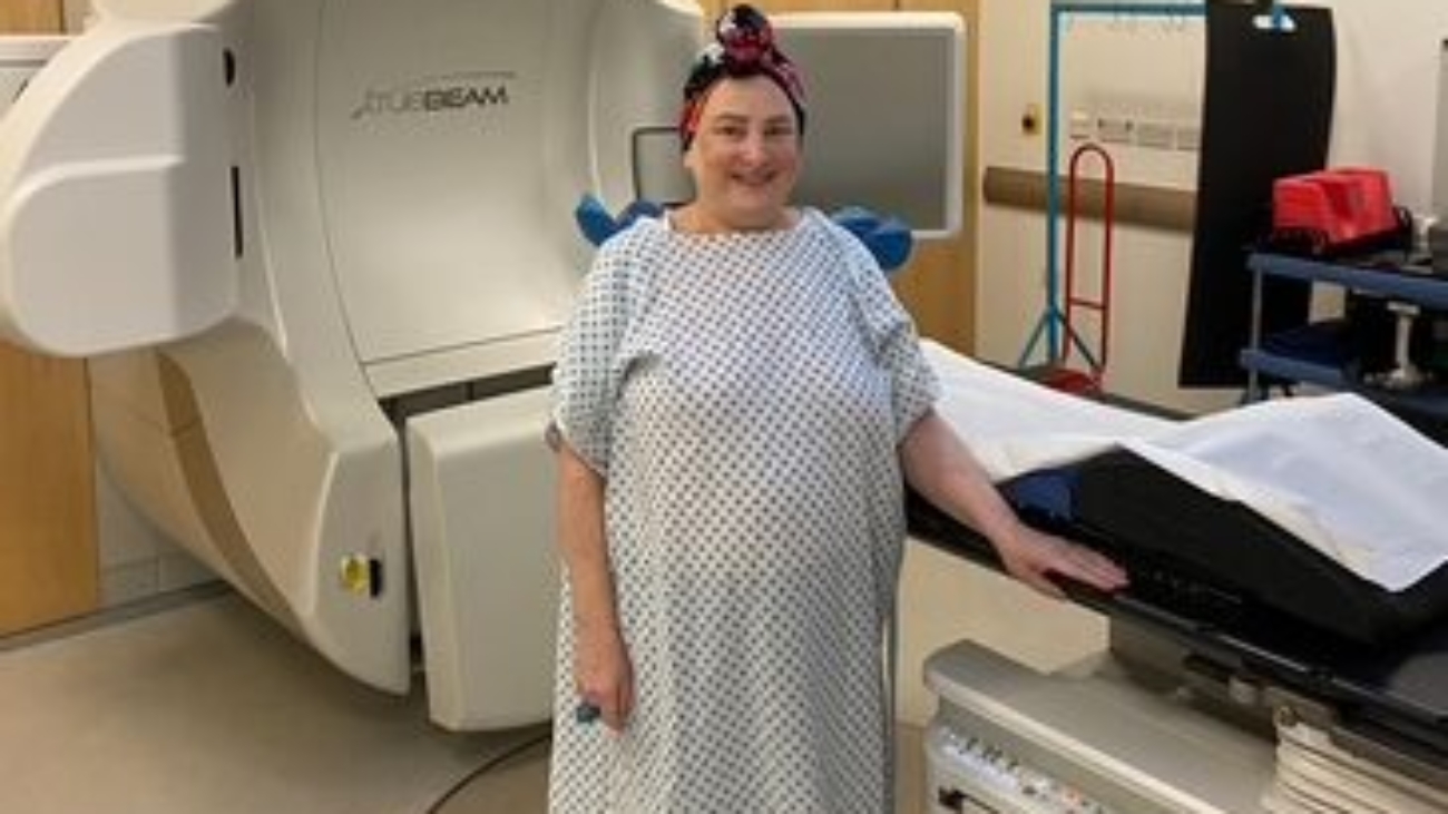
Helen at radiotherapy

Helen’s Breast Cancer Story

“That’s your treatment done”, best words I ever heard. I did a little dance to myself in the changing room!

In July 2022 I went for a regular mammogram… to my shock I was called back for further investigations and, the dreaded words  “confirming cancer” a couple of weeks later made the world stop for me. 

Then the roller coaster of tests tests and more tests,  and I felt in limbo until surgery then treatments started. I really was so very scared and I wish I could go back in time and tell myself, “all will be well”.

First came the surgery in August 2022, a lumpectomy carried out by Professor Zoe Winters. Her whole team made me feel relaxed and comfortable. I had a set back a couple of weeks later as I needed a second surgery but, again I was reassured this was very usual and, this time the margins were clear.

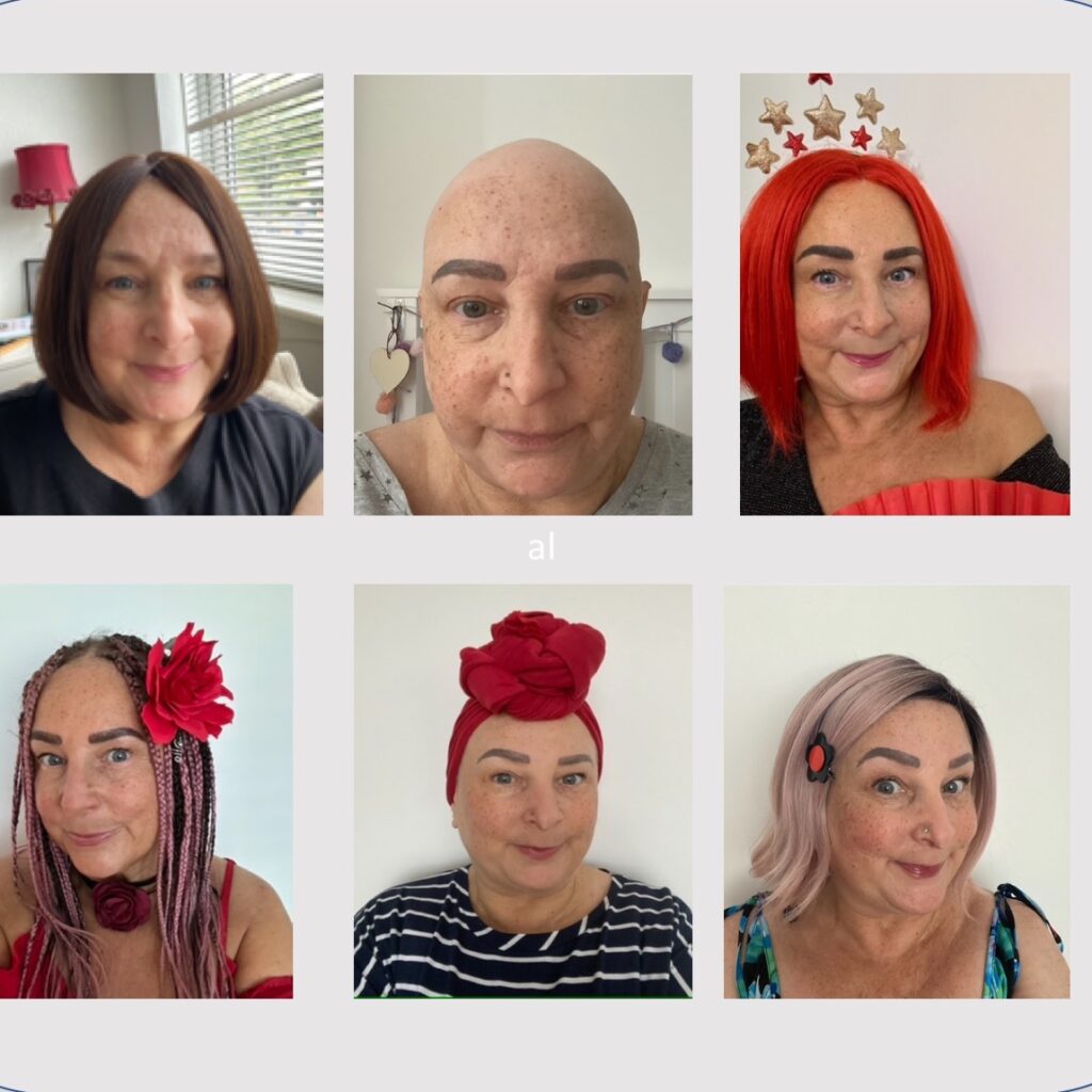
Next up, chemotherapy…. the team at the Royal Marsden were fantastic and when the day dawned of my first chemo session, although still terrified, it all went according to plan. The dreaded second chemo session meant my hair started to come out. Crying, I had to shave it all off, that was horrible. So, the only thing I could do, was treat myself to lovely pre-tied floral turbans and some exciting pink wigs! The options were endless. I became Etsy online stores best customer!
 
Pictures from normal hair to shaved hair, to wigs
Chemo was the hardest part for me. I imagined feeling nauseous all the time but I was fortunate to have such good anti-sickness drugs. I actually enjoyed going to the chemo lounge for my treatments, the patients were all so inspiring, chatty and from every walk of life, the nurses were also wonderful.

For me it was day 3 that hit the hardest after chemo. I couldn’t get out of bed I felt so weak and I had a rash and leg pain that were dealt with by the oncologists and nurses very promptly.  In total, I had 8 rounds of chemo and was so happy on the last one!

To my relief chemo was over. But then I had 15 radiotherapy sessions – every day! That really exhausted me.

On the last day of radiotherapy, the radiologist said, “That’s your treatment done”, best words I ever heard. I did a little dance to myself in the changing room! 

It takes a while to feel anything like yourself again! This was May 2023 and it took me until August 2023 to feel well enough to go back to my part time admin job. I felt so much better going back to work.

Helen after losing weight and final treatmentI had a girls holiday to spain in May 2024 and upon my return I went to see Professor Zoe for a follow-up. During the consultation Prof Zoe discussed my weight and referred me to a specialist dietician explaining that I had to lose the pounds because this may reduce my risks of the cancer returning.

I started my weight loss journey and am proud to say 16 months later, I am 4 stone (25.4 kilos) lighter!

I had reconstruction surgery in March this year (2025) and very pleased with the results.

My thin hair stayed thin and the medication I take to keep cancer at bay has possibly kept it thin? But, I have treated myself to lovely long tresses by a specialist clinic in Peterborough and am enjoying my hair and life again!

I am fortunate that with the excellent care I received, I have every chance that I will have the future I deserve. 

Ladies who are starting on their cancer journey, I just want to say, you will love your life again and please do not be afraid. I feel better than before I was diagnosed!
 
Helen x
Image showing HRT on a noticeboard and pills

HRT and Breast Cancer risks

More than 80% of breast cancers are due to lifestyle factors in the context of increasing age, clinical factors, and one’s individual population genetics which makes each one of us “different”, and is a consequence of multiple small changes in one’s DNA after birth.

We all experience a natural and progressive reduction in our circulating female sex hormones: oestrogen, progesterone and proportionate levels of androgens or male hormones. Progesterone levels are the first to decline at approximately 35 years of age. Our levels of oestrogen decline later around 45 to early 50s in the majority of women. 

Our female sex hormones are constantly changing within our body secondary to reproductive factors (age of menarche; age of first live birth, number of children and duration of breastfeeding). These inherent levels of natural hormones (called endogenous) within each woman may be affected by adding hormones 

(called exogenous) such as oral contraceptives and HRT.

Other lifestyle factors are likely to act through hormones such as: adiposity (amounts of body fat) and alcohol consumption. One’s physical activity is also an important modifiable risk factor. 

Endogenous (inherent individual) hormone levels are influenced by childbearing, which reduces breast cancer risk by 7% per birth. Breast feeding further reduces breast cancer risks by 4% per year of breastfeeding.

Exogenous (external addition) hormones such as oral contraceptives administered over 15 years at various ages have a very small increase in breast cancer risk that is greatly outweighed by a corresponding reduction in uterine and ovarian cancer, so that the net effect on female cancers is beneficial.

In a recent meta-analysis of 13 cohort studies in women under 55 years of age (median age 42 years) assessed over 8 years; 2% developed young onset breast cancer. 15% reported using hormone therapy; 6% used combined oestrogen and progesterone; 5% unopposed oestrogen. 4% of non-users developed breast cancer Lancet Oncology; 2025

Hormone therapy was not linked to incident breast cancer risk under 55 years of age. However, combined oestrogen (E) and progesterone (P), and use for more than 2 years, increased breast cancer risks (18%). This means an 18% higher risk of breast cancer occurring at any time point, compared to E only users or non-users. Combined E and P were associated with oestrogen negative (ER-) breast cancers, and triple negative cancers.  

Ovarian function ceases with menopause; thereafter oestrogen levels fall substantially and progesterone levels fall to near zero. Endogenous oestrogen synthesis in postmenopausal women occurs mainly in adipose/fatty tissue, catalysed by the enzyme aromatase. Postmenopausal oestrogen levels therefore correlate strongly with the amount of adipose tissue, and hence body mass index (BMI). Postmenopausal oestrogen is a strong determinant of ER+ breast cancer, whereby the latter accounts for three quarters of all breast cancer cases and deaths. 

In a meta-analysis of all studies on the duration and types of menopausal HRT used, and breast cancer risk www.thelancet.com, the authors conclude that there are excess breast cancer risks in women from 50-69 years of age, for women using HRT for 10 years and longer, starting at 50 years. This pertains largely to combination HRT (oestrogen and progesterone). The risk in these groups varies from 1 cancer in 25 users (continuous combined progesterone), to 1 cancer in 30 users (intermittent/cyclic combined progesterone). The risks are significantly lower in women taking oestrogen only (permissible only in those without a uterus, following a hysterectomy) where the risk is 1 extra cancer in 100 users. These risks persist up to 10 years in past users, after stopping HRT.

The above risks pertain to greater risks of: invasive cancers compared to never users; current HRT users; and persist up to 10 years after stopping HRT. The excess risks are greater for oestrogen receptor positive (ER+) cancers than for oestrogen receptor negative (ER-) disease. The risks are also greater for invasive lobular cancers compared to invasive ductal cancers. The breast cancer risks vary according to BMI, where increases in perimenopausal BMI are associated with increases in breast cancer risks in never users of HRT, that persists across all ages. The risks associated with increasing BMI are not seen amongst current users of HRT, as adiposity attenuates the excess risk of HRT. In obese women, the use of oestrogen-only HRT adds little to the adiposity-associated stimulation of their breast tissue. However, the risks are higher in lean women who are current users of oestrogen only HRT. Lean and obese women have similar absolute increased risk with the addition of progesterone.

Summary of this meta-analysis: For women of average weight in developed countries, 5 years of HRT use starting at 50 years of age would cause an appreciable increase in the probability of developing breast cancer at ages 50-69 years. 

However, it is important to note that the study is based on 24 prospective studies comprising three quarters of postmenopausal women (number = 108,647) that were diagnosed with invasive breast cancer at 65 years (mean age) in 2005 (median year). The 34 retrospective studies comprised a quarter of women (number = 35,240) diagnosed with invasive cancer in 1995. 

Current protagonists of HRT for menopausal symptoms and quality of life benefits discuss the fact that previous HRT preparations were synthetic, as opposed to being body- and bio-identical. There were likely issues of recall for women in retrospective studies, or in general relating to past use, and duration of use. 

So called “modern” HRT is underpinned by our understanding of the overall health and quality of life benefits of replacing oestrogen. I refer to this as the “queen of hormones” with reductions in: cardiovascular (heart) disease, dementia, abdominal obesity, and overall breast cancer mortality (deaths related to breast cancer). Oestrogen replacement alone does not increase breast cancer risks compared to the average population risks that are multifactorial in all women (breast cancer risk prediction tools). It is the addition of progesterone, or combined (oestrogen and progesterone) HRT that raises these risks. It is unclear why oestrogen-progesterone preparations would have a greater effect than oestrogen-only HRT.

In principle, modern HRT should comprise bio-identical oestrogen that is ideally administered through the skin as a gel or patch. The majority of women with an intact uterus require progesterone to stop the unopposed stimulation of oestrogen acting on the uterine lining to produce thickening thereof and breakthrough bleeding. Likewise, bio-identical progesterone is advised and should be used in the lowest doses in a sequential manner that is recommended by your menopause specialist or consultant gynaecologist. This may entail a combined oestrogen and progesterone skin patch, intra-uterine progesterone (Mirena coil), or an oral progesterone tablet called Utrogestan. Utrogestan contains micronized natural progesterone (called body-identical). It is derived from plants such as yam and soy, and is chemically identical to progesterone produced by the ovaries.

Current recommendations are to use bio-and body-identical HRT, and to consider type of administration in the context of individual breast cancer risk (breast cancer risk assessment tools). This is underpinned by contemporary breast screening individualised to mammographic breast tissue density and bespoke frequency of breast screening.

Breast cancer risks

There are a number of validated breast cancer risk assessment tools that can be referred to in Professor Zoe Winters’ blog on Young Onset Breast Cancer 13/06/2025

These algorithms are validated tools developed to assess the risks of carrying a BRCA 1 or 2 mutation, and of developing breast and ovarian cancer such as: BOADICEA/CanRisk or IBIS. They incorporate genetic results, family history, polygenic risk scores, clinical and lifestyle factors and hormonal data.

Women at high risk of developing breast cancer (estimated lifetime risk of 20% or more) may still consider HRT. However, each woman needs to understand the associated risks in the context of the Lancet 2019 meta-analysis, and the use of “modern” HRT that is carefully managed between their consultant breast and menopause specialist, and their geneticist. 

The risk assessment tool that I currently use is called the Breast Cancer Surveillance Consortium Risk Calculator that helps categorise women into low, intermediate and high risk, without integrating genetic testing. This guides my recommendations regarding breast screening, in terms of age of starting, and frequency intervals. Every woman undergoing a mammogram should know their breast tissue density, where dense breasts require additional imaging that includes IV contrast: either using a contrast enhanced mammogram (CE-M), or an abbreviated breast MRI This is based on the latest findings of the UK BRAID randomised trial featured in the Daily Mail 01/06/25.

Lifestyle factors

Body mass index (BMI): A BMI of 25 – 29 at 60 years of age increases breast cancer risks by 20% compared to a BMI under 25. This is also associated with increases in ER+ breast cancers compared to ER- cancers. 

Alcohol: Consuming more than 10g of alcohol per day increases risks of breast cancer that are equivalent to taking oestrogen only HRT, or 1 cancer per 100 women before 75 years of age. These are typically ER+ breast cancers. 

Breast tissue density: BRAID trial

Both these lifestyle factors are integrated into the CanRisk risk assessment tool, including the mammographic assessment of breast tissue density.

References: 

  1. Collaborative Group on Hormonal Factors in Breast Cancer. Lancet 2019; 394: 1159-1168.
  2. London Breast Health blog on Young Onset Breast Cancer, 13/06/2025
  3.  Hormone therapy use and young-onset breast cancer: a pooled analysis of prospective cohorts included in the Premenopausal Breast Cancer Collaborative Group. Lancet Oncology 2025; 26: 911-23
  4. Comparison of supplemental breast cancer imaging techniques—interim results from the BRAID randomised controlled trial. Lancet 2025; 405 (10493): 1935-1944
Young woman comforting her sad friend in the park during autumn

Young Onset Breast Cancer by Professor Zoe Winters. Part 1

Part 1, describes causes and risk factors on young onset breast cancer.

Young onset breast cancer is uncommon with 20 % of all breast cancers occurring under 50 years of age. Young age is defined as less than and equal to 45 years, but also includes women under 50 years of age. In the USA, 10-11% of women are diagnosed under 45 years of age, which is comparable to the UK. The greatest risks relate to women diagnosed under 40 years. More African American women are diagnosed before 50 years of age compared to Caucasian women. YOBC comprises only 6-10 % of all breast cancers in the developed world, compared to 20% in developing countries such as: East Asia, Middle East, North Africa and South America. Death rates vary from 7% in developed countries, to 14 % in developing countries. 

Young onset breast cancer in women is associated with special survivorship issues: fertility, contraception, early treatment induced menopause and pregnancy.

Women with young onset breast cancer are usually diagnosed with a clinical lump, and have more advanced clinical stages at diagnosis. The types of breast cancer are also more aggressive: such as triple negative (22%) and HER-2 positive (9%). These characteristics are assessed by taking a tissue biopsy of the cancer under image guidance (usually ultrasound). The tissue is analysed by the pathologist under the microscope to assess the type of breast cancer, histological grade or the appearance of the cells, the oestrogen receptor (ER), progesterone receptor (PR) and the HER-2 protein, plus cellular proliferation or ki67. These types of cancer (triple negative and HER-2 positive) present typically in women under 40 years of age. Generally, young onset cancers present with the following features: High histological grade (HG), which means they look more aggressive under the microscope, and they have a higher percentage of growing or dividing breast cancer cells measured as a snap-shot in time called a proliferation index (ki67). They harbour higher levels of the protein or oncogene called HER-2 or Epidermal Growth Factor-2 called HER-2 positive (HER-2 turns on the growth of cancer cells and is treated using drugs that block and turn off the HER-2 gene). Young onset cancers are also more likely to comprise triple negative cancers (TN), are oestrogen receptor (ER) negative (ER 0-3/8), progesterone receptor negative (PR) and HER2 negative on tissue staining under the microscope). The basal types of breast cancer express specific markers within the triple negative (TN) group: Epidermal growth factor receptor and markers called cytokeratins within the milk duct wall. This group is strongly associated with the BRCA 1 gene mutation. 

A large UK study called POSH in over 2000 women under 40 years of age; who were tested for, and shown to carry the BRCA gene, did better clinically in the short term up to 5 years, compared to the non-BRCA carriers. This may relate to being able to combine drugs that target the BRCA gene defect and enhance cancer cell killing (called PARP2 inhibitors), when combined with chemotherapy. The PARP2 drugs target and sensitize the cancer cells to be optimally killed by chemotherapy drugs, as they further reduce the ability of cancer cells to repair their DNA damage caused by chemotherapy. This pathway is already defective in BRCA gene mutated breast cancers. Beyond 5 years, the clinical outcomes were the same in women with or without a BRCA gene mutation over 8 years.

Of concern, are the increasing numbers of young women with luminal A types of breast cancer, which means that they grow under strong hormonal influence with strongly positive expressions of the oestrogen (ER), and the progesterone receptors (PR). The ER positive cancers are more likely in the BRCA 2 gene carriers in young women. The mechanisms underlying the development of these cancers is generally poorly understood with 90% showing no individual gene mutation, like BRCA1/2. Luminal B types are typically positive for the ER, but either positive or negative for the PR, and either positive or negative for the HER2 oncogene. These are more aggressive than the Luminal A types with higher cell proliferation and growth (ki67). Up to 20% of the Luminal B cancers are associated with HER2 positivity, which is responsible for these cancers being more aggressive. However, current new dual blocking drugs can turn off the HER2 growth pathway, and optimally kill cancer cells with excellent clinical outcomes.

What are the biological causes?

Germline or single gene changes (faulty gene in either the sperm or egg, that is transmitted to children) gene testing:

Mutations of genes can relate to specific inherited single genes that are passed on at the time of conception, either from one’s mother, father or both parents. Germline changes refer to the DNA you have in every cell you are born with.

This is called a hereditary germline gene pathway and is responsible for 10-20 % of young onset breast cancer. Clinical guidelines recommend genetic assessment for all patients with first-and second-degree relatives diagnosed with breast cancer younger than 50 years. 

High penetration genes (the penetrance of a disease-causing mutation is the proportion of individuals with the mutation that exhibit clinical symptoms among all individuals with such a mutation). Examples of these genes are: BRCA1, BRCA2 that are involved in repairing damaged DNA or genetic material. Only 10% of young onset breast cancer are shown to carry a BRCA1/2 mutation. In the UK POSH study, 12 % of women under 40 years of age were shown to carry a BRCA gene mutation. That said, BRCA 1 and 2 account for 50% of gene mutations in early onset breast cancer (they are referred to as the DNA repair genes). In addition, tumour suppressor genes that stop cancers from developing are: TP53, PTEN, STK11, and CDH1. These genes are responsible for 20% of hereditary risks. Ashkenazi Jews have a 10-fold increased risk of carrying a BRCA 1 or 2 mutation, and should undergo routine testing.

Moderate penetration genes are responsible for 5% of hereditary risks such as:

PALB2, BRIP1, ATM, CHEK2, RAD51C.

The above is responsible for only half of genetic risks, with the other half currently unknown. 

Therefore, it is recommended that the following 12 genes are tested in young onset breast cancers.

High-Penetrance Genes

  • BRCA1 and BRCA2: Found in 28.1% and 4.1% of very young patients (≤30 years), respectively. These genes confer high lifetime risks of breast, ovarian, and other cancers. Updated guidelines recommend testing all breast cancer patients ≤65 years.
  • TP53: Second most prevalent gene (4.5%) in patients ≤30 years, even without family history (74.8% of carriers lacked familial cancer history). Associated with Li-Fraumeni syndrome, warranting early inclusion in testing panels for this population.

Additional High- and Moderate-Penetrance Genes

Testing should extend to:

  • CHEK2, ATM, PALB2: Moderate-risk genes linked to increased breast cancer risk.
  • PTEN, CDH1, STK11: High-penetrance genes for syndromes like Cowden (PTEN) and hereditary diffuse gastric cancer (CDH1).
  • RAD51C, RAD51D, BARD1: Implicated in homologous recombination repair defects

Therefore, a blood or oral buccal mucosa (inner cheek scrape) test for assessing germline genes should be repeated every 6-8 years based on the American Society of Clinical Oncology (ASCO) guidelines. 

Currently, the NHS only tests 5 genes: BRCA1/2; PALB2; ATM and CHECK2, and potentially RAD51C and D where there is a history of ovarian cancer. 

Somatic mutations (faulty gene in the bodies normally functioning genes; these faults occur after conception and are not transmitted to one’s children): 

Somatic mutations are seen in cancer cells and cannot be inherited unless they are related to your germline. 80% of young onset breast cancer occur in-non carriers of a germline gene mutation.

Whole genome (DNA, specific gene code) or exome (the gene codes for and produces a message to produce a functioning protein) sequencing means reading abnormalities in the gene or the protein that detects abnormal gene and protein expressions that predisposes a cancer cell to develop. The earliest form of breast cancer develops from a normal breast duct epithelial cell: cells that line the wall of a microscopic breast milk duct, and is called ductal cancer in situ or DCIS. This mechanism for DCIS development involves deregulation of multiple gene and protein pathways that result in a cancer developing and growing, such as DCIS. 

The Molecular Taxonomy of Breast Cancer International Consortium (METABRIC) database showed high mutations in the CDH1 gene, that is responsible for encoding a key protein called E-Cadherin, that allows cells to adhere/stick to each other, and to the surrounding tissue called the extra-cellular matrix (tissue that surrounds the microscopic breast ducts). Cell to cell adhesion is reduced when a cancer develops. When these pathways are abnormal, E-cadherin levels go down, and cancer cells spread/detach or metastasize, leading to a poor prognosis. An e.g., of a particular type of cancer that results from this CDH1 gene mutation is the invasive lobular cancer or ILC, that spreads in a single microscopic file along the breast milk ducts. This is referred to by your surgeon as a ‘single file pattern’.

Other important genes:

GATA3 is important in developing the breast gland tissue, and in maturing the immune cells, called T cells. T cells are part of the immune system and develop from stem/originator cells in the bone marrow. They help protect the body from infection and may help fight cancer. They are also called T lymphocytes. Cancers that are defective in GATA3 have poorer outcomes, with shorter overall survival.

CTNNB1 is a gene that encodes Beta-catenin that also controls cell to cell adhesion (how cells stick together). If these genes don’t function and the levels of catenin drop, then cancer cells spread and don’t respond to anti-oestrogen hormone blockers like tamoxifen or letrozole.

TP53 (cancer suppressor gene that detects DNA/cell damage) and PIK3CA (growth factor pathway on the cancer cell surface) mutations are seen in up to 40% of cancers.     

Family history of Young Onset Breast Cancer:

Clinical guidelines recommend genetic testing for all women with first- and second-degree relatives with young onset breast cancer, occurring younger than 50 years of age.

A risk prediction tool called CanRisk is advised to estimate the predicted risk of a patient carrying a disease producing gene for either breast and/or ovarian cancer. If this estimate is 10%, a referral should be made for genetic testing. There is no consensus to support the recommendation of one risk prediction model only. Most tools predict 5- and 10-year risks of developing breast cancer, but not how young this will occur. 

This is called testing for a possible inheritable germline gene inheritance. A germline gene refers to a gene present in reproductive cells (sperm or eggs) that can be passed down to offspring, meaning any changes or mutations in these genes can be inherited by future generations. Living relatives with index cancers should be tested first using an appropriately large/extended gene panel to reflect their cancer and other cancer types that may link to breast in family members. Each sibling in a high-risk family should be tested as the potential risk of inheritance is 50/50. This approach to initial blood testing should always be discussed with a recognised genetic counsellor within a recognised genetics centre of excellence.

Presently, studies provide evidence to support the risk-reducing benefit of offering risk-reducing surgery (breasts and/or ovaries) in all patients shown to have mutations in: BRCA1, BRCA2, PTEN, and P53. Regarding all other gene mutations, the patient’s individual risks should be considered by using an evidence-based algorithm called CanRisk (https://www.genomicseducation.hee.nhs.uk/genotes/knowledge-hub/canrisk/). It is important to refer each patient for a consultation with a geneticist. It is important to select the appropriate and most extended gene panel (which means increasing the number of genes tested). Each patient needs to understand the numbers of genes that have been tested and that the results may need to be reassessed 6 yearly, as the scientists discover new genes. 

Single nucleotide polymorphisms (SNPs) and polygenic risk scores (PRS) explained:

The body’s genetic code is contained within one’s DNA that comprises a helix or twisted ladder containing multiple rungs comprising sugar base pairs represented by pairing sugars. When there is a single base or sugar letter change in a rung of the ladder, this is called a single nucleotide polymorphism (SNP), that on its own can cause a very small increased risk of developing a disease. However, when multiple SNPs and their individual risks are combined, there is an increased cancer polygenic risk score (PRS) risk for breast cancer. Therefore, the genetic team needs to test for a single germline gene and polygenic risk scores (PGRS).

The breast cancer risk prediction tool that uses polygenic risk scores (PRS) is BOADICEA, which is accessible through the CanRisk webtool. Breast and Ovarian Analysis of Disease Incidence and Carrier Estimation Algorithm (BOADICEA version 5) incorporates PRS alongside other genetic and non-genetic factors, such as family history, mammographic density, and lifestyle factors, to provide comprehensive risk assessments for breast and ovarian cancer.

BOADICEA’s inclusion of PRS enhances its predictive accuracy and ability to stratify individuals into different risk categories for personalized prevention and screening strategies.

Risk Assessment tools: 

Current recommendations are to identify women at risk of familial cancer syndromes based on a detailed family history. Validated tools were developed to assess the risks of carrying a BRCA 1 or 2 mutation, such as BOADICEA/CanRisk or IBIS. Guidelines for genetic testing recommend testing no earlier than 18 years of age. 

The CanRisk breast cancer prediction tool includes BOADICEA. BOADICEA stands for Breast and Ovarian Analysis of Disease Incidence and Carrier Estimation Algorithm, which serves as the foundational model for CanRisk. It integrates genetic factors, family history, lifestyle, hormonal/reproductive factors, polygenic risk scores, and mammographic density to estimate breast and ovarian cancer risks.

Validated models are used to assess estimated cancer risks at 5 and 10 years, including lifetime risk of breast cancer, such as the Breast Cancer Surveillance Consortium (BCSC)

These are not necessarily specific to assess the risk of early onset or premenopausal breast cancer. The choice of tool depends on individual patient characteristics and clinical needs.

1. BOADICEA/CanRisk (Breast and Ovarian Analysis of Disease Incidence and Carrier Estimation Algorithm)

  • Incorporates genetic factors (e.g., BRCA mutations), family history, and polygenic risk scores (PRS).
  • Available through the CanRisk webtool, it is highly accurate for both breast and ovarian cancer risk estimation.

2. IBIS (Tyrer-Cuzick Model, women older than 20 years) 

  • Combines genetic information, family history, lifestyle factors, and hormonal data.
  • Recently updated to include PRS for enhanced predictive accuracy.

3. Breast Cancer Surveillance Consortium (BCSC) Risk Calculator (women older than 35 years)

  • Includes mammographic density alongside personal and family health history to estimate risk.
  • Particularly useful for integrating radiographic factors into risk prediction.

Table 1: Risk factors included in CanRisk estimates

Risk factor

Breast cancer

Ovarian cancer

Family and personal proband history of breast, ovarian, prostate and pancreatic cancer

High-risk constitutional (germline) pathogenic variant

BRCA1, BRCA2, PALB2, CHEK2, ATM,

RAD51D, RAD51C,

BARD1

BRCA1, BRCA2,

PALB2, RAD51D, RAD51C, BRIP1

Age of family members (affected and unaffected)

Ashkenazi Jewish heritage

Year of birth (birth cohort)

Age at menarche


Parity

Age at first birth


Oral contraception use

Hormone replacement therapy use

Body mass index

Height

Alcohol use


Mammogram breast density


History of tubal ligation


History of endometriosis


Polygenic risk score

If a woman’s estimated lifetime risk of developing breast cancer is 20% or more, one is regarded as high risk. This merits early breast screening from 30 years of age. The USA National Comprehensive Cancer Network recommends starting yearly mammograms at 30 years, and yearly breast MRI at 25 years.

Other predisposing risk factors:

Dense breasts: The fibro glandular breast duct tissue appears white or brighter on the mammogram compared to darker fatty tissue. Dense breasts are present in the majority of younger women under 46 years of age. This is a subjective assessment about how much fibro glandular tissue there is. The highest density raises the risk of breast cancer 4-6-fold. However, most of this data comes from women older than 50 years of age.

High density on mammograms lowers their sensitivity (reduced ability to detect a cancer) to 62%, from 88%. This can result in more interval cancers, that means a cancer develops in between recommended screening intervals, after reporting a normal mammogram. Interval cancers are increased 2-fold in dense breasts and have a worse prognosis, based on their larger size, and higher lymph node positivity. 

Decisions recommending additional screening using ultrasound, should consider additional risk factors, and are indicated, where individual risk is above average.

Proliferative Breast disease: We list benign breast changes that have either increased risk of being associated with DCIS (Ductal Cancer in Situ), which means microscopic breast cancer cells that are contained within microscopic breast ducts, or predict increased risks of invasive breast cancer developing. 

    1. Atypical ductal hyperplasia (ADH): carries a 20% risk of associated DCIS or invasive cancer. This lesion requires diagnostic excision
    2. Radial scar: carries at 8-15% risk of associated DCIS or invasive cancer. This lesion requires diagnostic excision 
    3. Atypical lobular hyperplasia (ALH) or ADH: carries a 4-fold increased lifetime risk of breast cancer 
  • Lobular carcinoma in situ (LCIS): increases the lifetime risk of breast cancer over 10 years to 7%
  1. Pleomorphic LCIS: further increases the lifetime risk of breast cancer, and is an aggressive form of LCIS. It requires surgical excision

These lesions predict higher breast cancer risks and should be discussed with a Consultant Breast Specialist. Although data is lacking, increased breast screening is advised.

  1. Yearly mammograms and tomosynthesis 
  2. Yearly breast MRIs
  3. Clinical examination yearly 

Hormonal contraception:

Current oral contraceptive use poses small increased risks of breast cancer (1 extra cancer per 7700 women). The oral contraceptive pill significantly reduces risks of uterine and ovarian cancer. There are no specific breast screening guidelines in this context.  Intrauterine or injectable progesterone poses no increased risks.

Fertility Treatments:

There is no conclusive evidence that fertility drugs are associated with increased risks of breast cancer. The American Society of Reproductive Medicine states that there is “fair evidence that fertility drugs are not associated with increased risk of breast cancer”. Therefore, routine breast screening is recommended in the absence of other risk factors.

History of radiation exposure:

Radiation treatment to the chest wall before 30 years of age is a high-risk factor for EOBC. These treatments may comprise “mantle radiotherapy” for Hodgkin’s lymphoma, with the greatest risks in women treated with 40 Gy or more. However, lower or moderate dose chest radiation of greater than 20 Gy also increases risks. The risks are equivalent to having a BRCA 1 or 2 mutation, comprising a 13-20% risk of EOBC by 40-45 years. These women should be offered high risk breast screening.

Prior Breast or Ovarian cancer: 

Among young onset breast cancer survivors, the risks of a second early onset breast cancer are unknown, unless there is a high-risk gene mutation, like BRCA 1 or 2. There is no data for the risk of early onset breast cancer in women with early onset ovarian cancer. Yearly mammograms are recommended at 6-12 months after completion of treatment.

Other links to related blogs by Professor Zoe Winters.

  1. Personalised genetic testing in breast cancer patients and what it means.
  2. Breast density 
  3. Breast feeding 

References:

  1. Chelmow D et al. Executive summary of early onset breast cancer evidence review committee. Obstetrics and Gynaecology 2020, 135 (6): 1457-78.
  2. Gao Y et al. Non-BRCA early onset breast cancer in young women. Radiographics 2022; 42: 5-22
  3. Copson ER et al. Germline BRCA mutation in young onset breast cancer (POSH): a prospective cohort study. Lancet Oncology 2018;19:169-80.

London Genetic Centre: https://www.thelondongeneticscentre.com/

General template 3

Breast Cancer Surgeon Professor Zoe Winters Emphasises Clinical Evidence to Reduce Recurrence Following Elle Macpherson Cancer Diagnosis

Recent news reports have Super model, Elle Macpherson published that Elle Macpherson has revealed that she refused medical treatment following a cancer diagnosis.


After undergoing a lumpectomy to remove cancerous tissues, Elle was diagnosed with HER2-positive oestrogen-receptive intraductal carcinoma – a type of breast cancer and was subsequently advised by doctors to undergo a mastectomy with radiation, chemotherapy, hormone therapy and a breast reconstruction for treatment. However, in February 2017, the supermodel mum of two decided to refuse medical treatment and chose a holistic approach to cancer therapy.

After her diagnosis, Elle spent eight months in Phoenix, Arizona, where she was supported by her personal doctor, holistic dentist, osteopath, chiropractor, naturopath and two therapists. During this time, she focused on “addressing emotional as well as physical factors associated with breast cancer”.

Elle has since reported that at 60 years of age, she is now in “clinical remission” after being diagnosed seven years ago.

Following this news, we asked Professor Zoe Winters, Professor of Breast Cancer Surgery at University College London Hospital and New Victoria Hospital in a Q&A to provide insight on the diagnosis and highlight the impact of medical intervention vs alternative holistic therapy.

Detailed Q&A below…

1. Ms Macpherson was diagnosed with HER2-positive oestrogen-receptive intraductal carcinoma – a type of breast cancer – after undergoing a lumpectomy to remove cancerous tissues.  Can you explain this type of cancer?

Intraductal cancer is defined as Clinical stage 0 breast cancer, which is mostly referred to as Ductal Cancer in Situ or DCIS. The generalised term “cancer” is vague as DCIS by definition comprises microscopic breast cancer cells that are contained within a microscopic breast duct.

DCIS is a predictor of bilateral increased breast cancer risks, however, this refers to high risk within the average population and is not equivalent to very high risks that relate to an inheritable gene mutation such as the well-known BRCA genes.

HER2-positive DCIS does not equate to the poorer prognostic impact of HER2-positive invasive ductal cancer where breast cancer cells have invaded outside a microscopic duct and therefore may have spread to axillary lymph nodes.

However, it is important to note that the information shared in news reports on the case of Elle MacPherson is vague, with few clinical details that help clinicians or patients to interpret their possible risk.

2.  In light of this diagnosis, what options would a patient be offered for treatment?

As a way of assessing risk, population risks are not an indication that bilateral risk-reducing mastectomies are required or beneficial, however, oncologists offer women the opportunity to consider chemo-prevention (medical risk-reducing) treatments using drugs like Tamoxifen or aromatase inhibitors for 5 years, based on international randomised (studies designed to reduce bias) clinical trials based on 10 of thousands of women. This is called level 1 or the highest quality of clinical evidence that allows clinicians to recommend a particular treatment.

3. What factors influence the risk of Elle Macpherson’s cancer?

Several factors influence the future risks of DCIS such as young age of onset or less than and equal to 50 years, large size of the DCIS, whether the DCIS presents as a lump, and whether a wide local excision or surgery lumpectomy has clear microscopic margins that should measure at least 1-2 mm away from the edges of the excised surgery specimen or wide local excision.

4. What do you advise patients who decide not to go ahead with any medical treatment and what is the potential outcome for those who refuse treatment?

HER2-positive DCIS has been studied in a large UK/ANZ DCIS trial in over 700 women where HER2 expression predicts a significantly increased risk of DCIS recurrence, and also strongly predicts radiotherapy benefit after wide local excision, with a greater reduction in DCIS recurring in the same breast.

5. Do you have any other advice for women considering alternative routes to cancer therapy?

Modern medicine is constantly evolving where clinical evidence is strengthened by designing studies that limit bias, that may exist by treating clinicians and patients. Bias is a perception not based on the body or strength of existing evidence.

Lifestyle factors are important in improving overall health but are not considered clinically equivalent to proven treatments based on studies that are numerically powered to show a difference in clinical outcomes when a particular treatment is used.

Evidence-based studies account for random and placebo effects in medicine on both sides of the consulting desk.

About New Victoria Hospital

New Victoria Hospital is a private, charity-owned hospital based in Kingston upon Thames, Surrey and is one of the few remaining independent hospitals in the country.

It has provided a high level of service to the local community for over sixty years and is frequently ranked by patients as one of the top private hospitals in London.  The Hospital is registered with and regulated by the Care Quality Commission (CQC) and is fully compliant with their standards.  The current CQC rating for the Hospital is “Good”. The. The hospital was proud to achieve this rating across all five categories–safe, effective, caring, responsive and well-led. It is owned by parent charity The Victoria Foundation. This charity helps to transform lives where there is an opportunity to do so either through medical provision or by ensuring that young people destined to become future generations of doctors are not prevented from doing so through lack of finances.

Article here – CLICK HERE

Cancer patient 6 months on from diagnosis

Del’s Breast Cancer Story

18th January 2024 – A DAY OF CELEBRATION – My ‘little’ brother is 46 today, and my oncologist has told me following cancer treatment and two spine biopsies, I have no visible cancer in my body 

It came in three’s

Hello, my name’s Deljinder. I’m 51 and on the 7th July 2023, following my first routine NHS screening, a lump was found and I was required to undergo a biopsy. From here I was diagnosed with breast cancer.

Following a PET CT and MRI in late July, I took a call telling me a suspicious lesion had now been found on my spine.

One must soldier on….

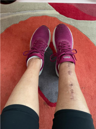
On the 9th September (my birthday) and 4 days before my planned surgery of a lumpectomy under Professor Zoe Winters, I broke my leg. A severe break resulting in a plate and 12 screws. Not being able to walk, surgery had to be cancelled and a later date was set.

I was lucky to have Professor Zoe Winters on my case, what an amazing woman.

Following breast cancer surgery, I recuperated at my mother’s which gave me lots of time to think..

Not one to feel sorry for myself, this thinking resulted in how I could help a Breast Cancer charity. So, I decided to sign up for the Moonwalk 2024 in Edinburgh. – challenge on. The walk is almost a year to the day that I broke my leg and was diagnosed with breast cancer.

I am hopeful that the combination of the walk and the charity will help with not only getting my physical strength back, but assist me getting back to me!

Every woman has their own strengths and being diagnosed with breast cancer certainly challenges our strengths and weaknesses!

I don’t hate the fact that the cancer picked me, I love the strength it has given me.

Powering on…. 

I hope this helps other women find their inner strength.

Picture of Del's new trainers to train for the Moonwalk and scar from broken leg

Mander’s Mammaries – MoonWalk challenge to raise £2,500 by 1st Sept 2024

A highly personal challenge following my breast cancer diagnosis and broken leg. Really excited to be walking as a duo team with Laura for The Moonwalk Scotland 2024!

                                                                                                Del’s moonwalkscotland2024/fundraising
 
Breast imaging report with jargon

Imaging report – understanding your mammogram and ultrasound report

A Consultant Radiologist will provide you and your Consultant Surgeon with an imaging report with numbered categories for each type of imaging. 

At each stage of the One-Stop triple assessment, the suspicion for malignancy is graded to create an overall risk index, as described below. The key below is used to establish whether a lesion is likely to be a benign, indeterminate, or highly suspicious that requires further imaging and or an image guided core needle biopsy.

(P) = Physical / Clinical examination by the Surgeon (P1, P2, P3, P4)

(M) = Mammographic Grade Description (M1, M2, M3, M4)

(U) = Ultrasound Grade Description (U1, U2, U3, U4)

(B) = Biopsy Grade Description (B1, B2, B3, B4)

What the codes mean on the report:

P1 / M1 / U1 / B1 = Normal 

P2 / M2 / U2 / B2 = Benign 

P3 / M3 / U3 / B3 = Uncertain / probably benign

P4 / M4 / U4 / B4 = Suspicious of malignancy 

P5 / M5 / U5 / B5 = Highly suspicious of malignancy

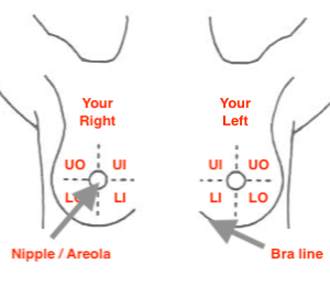
Quadrants: Right & Left

Each breast is divided into 4 quadrants as the surgeon faces the patient with the nipple areola in the centre. The right breast correlates with the client’s right, and the left breast with the client’s left. The Radiologist and Surgeon each describe the quadrant of the breast where the lesion is seen, whether it is central or peripheral, and also include location details by referring to the numbers of the face of a clock as one is looking at it.  

Picture showing a diagram of the breast in terms of radiology reports

UO: Upper Outer (superior and lateral)

UI: Upper Inner (superior and medial)

LO: Lower Outer (inferior and lateral)

LI: Lower Inner (inferior and medial)

C: Nipple and Areola (Central with clock numbered position)

IMF: Infra-mammary fold or bra line (inferior central, inferior lateral, and inferior medial)

Terminology found in your imaging report(s):

Addendum = The supplementary text added at the end of a previously approved radiology report, to correct or expand on an original assessment by the Consultant Radiologist. Additional radiology review is required where there is a need to compare current images with previous mammogram or ultrasound images.

Architectural distortion = This suggests a suspicious or highly suspicious lesion on a mammogram that may require a tissue biopsy, but can also be caused by scar tissue after breast surgery. Occasionally, a benign lesion such as a radial scar comprises benign breast ducts (adenosis) with extensions like spicules associated with fibrosis (scarring) that is normal tissue in more than 90% of cases.

Asymmetric density = A mammogram lesion seen in one breast compared to the same location in the other breast, that may require further investigations, if it is new compared to previous imaging. This may also arise from differences in the amounts of breast duct tissues between the breasts that are part of developmental differences at birth, and usually requires additional imaging views using magnification mammogram and 3 D mammograms called tomosynthesis.

Axillae = Armpits

Benign = Non-cancerous

Bilateral = Both breasts

Calcifications = Evidence of coarse calcium deposits that develop in normal breast tissue such as cysts, and benign fibroadenomas, including blood vessels and breast duct. Calcifications may also occur in malignant lumps, if slow growing, or if fast growing following death of breast cancer cells called necrosis. 

Complex breast cyst = Contains a solid tissue or polyp-like  growth within the cyst wall, and requires a tissue biopsy.

Complicated breast cyst = This type of cyst has some semi-solid particles / fragments floating in the fluid on ultrasound, and requires either aspiration, or a core tissue biopsy if the aspirate is either “bloody”, or the cyst doesn’t disappear. A complicated cyst may also be called a glue cyst (see below). 

A complicated cyst wall may also be thickened, and require a core tissue biopsy and not simply an aspiration. 

Cortical thickness = The normal thickness for the lymph node cortex is 3 mm. If the lymph node cortex is thicker, it requires a tissue core biopsy to exclude abnormal cells. Often the thickened lymph node is a normal reactive node that represents a normal immune response after a biopsy or infection. However, it is important to exclude cancer cells. 

Cyst = A cyst is a small cavity filled with fluid. Cysts are normal and arise from microscopic breast ducts as they age. Hormones like Oestrogen, Progesterone and Prolactin (milk producing hormone) can increase secretions by the breast duct cells that line the walls of the microscopic breast ducts. The breast duct walls become thinner with age causing the ducts to dilate called duct ectasia. The thin duct blows-out fluid filled cysts.  

Cysts are mostly described as benign and are rarely associated with cancer. Most cysts resolve spontaneously, and are nothing to worry about. If the cyst is large or causing discomfort, your Radiologist may draw off the fluid using a fine needle and a syringe, called cyst aspiration.

Fibroadenoma = A benign (not cancerous) developmental lump that occurs mostly in puberty and is not a risk factor for breast cancer. Fibroadenomas do not become cancers. They have an unknown cause but comprise a mixture of fibrous and breast duct or glandular tissue. They may fluctuate in size with hormonal triggers like the pill, hormone replacement therapy, pregnancy and breastfeeding. One third increases in size, one third becomes smaller, and one third disappears. They do not require surgery removal; however, surgery excision may be offered as a choice if they increase in size to over 30 mm. 

Fibrocystic changes = Normal breast duct tissues, admixed with fibrous connective tissues, and benign simple cysts.

Fibroglandular density = Normal overlapping breast duct tissues and fibrous tissue producing this appearance on the mammogram.

Findings = What was “found” from the radiology exam, listing each area of the breast that was examined in the diagnostic imaging study

Focal = This describes a change or finding in one area of the breast on imaging.

Glandular = Glandular tissue is another term for dense (increased breast duct tissue versus fatty tissue), normal breast duct tissue that appears white on mammogram and ultrasound. Greater amounts of glandular tissue occur in the upper outer breast areas, central nipple area, compared to the lower breast segments.

On clinical examination, glandular areas are firmer, compared to the fatty breast tissues that are softer.  

Glue cysts = Contains semi-solid particles. It requires cyst aspiration to determine if it disappears completely in which case, it is a simple or normal and benign. 

Hypoechoic mass = This means a lesion that is more dense or solid than usual as seen on an ultrasound scan. Ultrasound uses sound waves that are either absorbed (look black like a cyst which is fluid), or bounce off tissues (look white like a solid lump), based on their density. The sound waves form the black and white image you see on an ultrasound screen e.g. The whiter image is the breast duct tissue, and the darker image is fatty tissue or fluid that looks like a “black hole” in the case of simple cysts. 

Indeterminate = Atypical 

Indeterminate lymph node = The axillary lymph node may be larger compared to the other lymph nodes. The lymph node comprises a fatty centre surrounded by a rim of normal lymphatic cells called lymphocytes,that form a rim called a cortex. An indeterminate node may have a slightly thickened cortex that requires a lymph node biopsy if its thickness is greater than 3 mm.

Lipoma = This is a benign developmental growth of normal fat cells that forms a soft tissue benign lump with a defined capsule or border seen on ultrasound. They can be surgically removed if troublesome or increase in size, but lipomas in the breast do not increase breast cancer risks. 

Large lipomas may require MRI soft tissue imaging to ensure that they are not malignant sarcomas called liposarcomas. The latter should be referred to a specialist Sarcoma clinic. 

Lobulated = This refers to a benign fibroadenoma that has a smooth border with undulations called “lobules” on imaging.

Macrocalcifications = Appear as large white coarse spots that look like dashes or dots. They are rarely indicative of a cancer, and their presence does not require additional tissue biopsy testing or follow up, except routine breast screening.

Malignant = Cancerous

Microcalcifications = Appear as tiny finely clustered white specks. They are often referred to as appearing like “salt and pepper” on a mammogram. They are also usually non-cancerous, but if these tiny specks form certain patterns or have increased over time based on comparison mammograms, they can indicate a microscopic cancer that is confined to a microscopic duct called ductal cancer in situ (DCIS). Generally, further imaging, and evaluation is required using additional mammogram views and possible mammogram guided tissue biopsy.

Modified WHO criteria used = Form signed to say that allergies have been checked.

Nodular fibroglandular = This is described above under “Nodular Pattern”.

Nodular pattern = The normal breast is composed of dense fibrocystic breast duct tissues, that are intermixed with fatty tissues in a fairly irregular pattern that causes a nodular appearance on imaging, or a “lumpy” feel on clinical examination. Mostly, these are related to solid masses that appear white on mammograms and ultrasound in keeping with dense normal ductal breast tissue. 

Fibrocystic changes that contain sub-centimetre cysts are diagnosed on ultrasound as multiple “small black holes” in keeping with cysts that are normal.  

No focal solid masses = Normal breast tissues. 

Palpable lump = A palpable lesion / lump in the breast is usually subjected to triple assessment (Clinical examination, Imaging and Core Biopsy or Fine Needle Aspiration).

Partially cystic mass = This means a semi-solid lesion that has mixed solid and fluid components. This may suggest a complex cyst that requires a tissue biopsy of the solid component if the cyst doesn’t collapse completely on fluid aspiration, of the cyst fluid is “bloody”.

Sarcoma = Rare form of rapidly growing soft tissue cancer that may arise after radiotherapy for breast cancer treatment, or is a malignant phyllode tumour, that is a variant fibroadenoma diagnosed on tissue core biopsy or following surgery excision. 

Scattered microcysts = Less than 10mm in size. Too small to feel, and detected using ultrasound.

Scattered simple cysts = Cysts that can be felt are called symptomatic, and may be one or multiple in number. A breast cyst often feels like a mobile smooth grape, or a water-filled balloon, but sometimes feels hard. Breast cysts don’t require treatment, unless they are large and painful, which requires aspiration.

Sebaceous cyst = This is a benign developmental skin cyst that occurs in the dermis of the skin. It is not related to breast duct tissues. If troublesome, it requires a referral to a  Consultant Dermatologist for skin ellipse excision under local anaesthetic. 

Sub-centimetre cysts = Less than 10mm in size. These are not large enough to be seen on a mammogram, but are detected on ultrasound. They are benign and occur as part of duct ageing and breast duct secretions. 

Subcutaneous (cystic lesion) = See sebaceous cyst.

Superficially = This means close to the skin and fatty tissues underlying the skin.

Tomosynthesis or 3-D Mammogram = Advanced form of mammography that uses a low-dose x-ray system and computer reconstruction to create three-dimensional images of the breasts. Tomosynthesis is used for further evaluation or very dense breasts, and is considered to be enhanced breast screening.

Unifocal = This means a single lesion. 

Unilateral = Single breast

Unremarkable = If an area appears as normal breast tissue (there is nothing to worry about)

Vascular mass = This means lesion with increased blood flow seen on doppler ultrasound.

BI-RADS Mammogram ScoringUnderstanding your BI-RADS Score 

BI-RADS stands for Breast Imaging Reporting and Data System. It is an assessment tool used to rate the results of a mammogram test.

The BI-RADS score has seven levels of ranking:

  • Category 0: Additional imaging is required to provide a category. The results were inconclusive.
  • Category 1: Your test result is negative. There is no significant or noticeable abnormality on your mammogram. It is important to continue recommended routine screening.
  • Category 2: A benign non-cancer lesion was identified. There is a benign calcification or fibroadenoma, but it is not of concern. A tissue core biopsy may be indicated.  
  • Category 3: A lesion was identified that is most likely benign. You may require a tissue core biopsy if the lesion is solid or a follow up mammogram at 6 months to monitor the area for changes.
  • Category 4: There is a suspicious abnormality that may be cancerous. This result requires a tissue core biopsy.
  • Category 5: The growth is highly likely to be malignant, meaning it is probably cancer. This result requires a tissue core biopsy. ‌
  • Category 6: A biopsy confirms the results of your mammogram, and the growth is identified as a cancer. Treatment should begin and be directed by a Consultant Breast Surgeon and Breast Specialist multi-disciplinary team.

BI-RADS reporting mammogram breast density 

Your mammogram report will also include an assessment of your breast density, that describes the ratio between the amount of normal breast duct tissues and fibrous tissue called glandular tissues (whiter), compared to the fatty tissues (darker). The denser your breasts, the harder it can be to see abnormal areas on mammograms such as solid lesions, that can also appear white, making it difficult to separate a ‘white cancer” from normal dense breast duct tissues. 

In a mammogram report, the radiologist uses a standard system called BI-RADS or ACR (American College of Radiology).  The breast tissue density is classified into 4 categories from A-D. These categories describe the amounts of fatty tissues (seen as darker), compared to dense breast duct tissues (seen as whiter) in your breasts. The amount of breast tissue density increases from A to D, with D representing extreme breast tissue density as follows:

A: The breasts are almost entirely fatty with a greater proportion of darker tissues versus breast duct tissues that are whiter (about 10 % of the screening population). 

B: The breast comprises mostly fatty tissue with a few scattered areas of dense fibrous and glandular breast duct tissues (about 42% of the screening population). 

C: The breasts comprise multiple areas of fatty and dense breast duct tissues, that are referred to as heterogeneously dense, and may obscure small masses (about 40% of the screening population). 

D: The breast is almost entirely comprised of dense breast duct tissues, and is referred to as extremely dense, that lowers the sensitivity of mammograms (about 8% of the screening population). 

Cancer patient with her children

Martine’s Story

There is a saying that tough times never last, but tough people do, so when cancer touched my boob, I decided to kick its a*s!…”
 
Here is what I’ve learned so far 

– Cancer causes shopping – seriously, it’s one of the most dangerous side effects.

– At the beginning of your journey you will spend way more time making other people feel better about your diagnosis than them comforting you. Definitely stops you wallowing.

– Taking up yoga, meditating, burning candles and drinking green juice is not for everyone. Mostly made me want to smack some people (although the green juice is good). 

– The music on your Healthcare providers authorisation line will become the music you remember – hope it’s not 80s elevator music like mine.

– My godsons have coined a new phrase “Cancer Christmas” – it’s the festive period when all the people who love you send chocolate, brownies, fudge, cupcakes, hampers, cheese plates and your neighbours give you a LOT of lasagne. (Side effect –  no longer fitting into your new mastectomy bra).
 
Martine at Christmas with her boys
 
– Trying not to laugh when your seven year old tells you the doctor forgot to put your nipple back on is a difficult, but not impossible, feat.
 
And the last trite but true for me saying – the sun never stops shining, sometimes the clouds just get in the way… and any cancer diagnosis is more like an eclipse than a rain cloud.  But the sun is still there and every day throughout my treatment with the amazing Professor Zoe Winters and her team, it shines a whole lot brighter and the sky becomes clearer.  
 
I think a cancer diagnosis inevitably changes you, and I’ve decided it’s changing my life for the better.  I’m thankful everyday for my friends, family and loved ones who have done and continue to do so much for me. I’m so grateful to all the amazing medical professionals that are guiding me through my journey, but also to those behind the scenes that have discovered cutting edge medical treatments and cures, and help us fight this battle with such an incredible arsenal of weapons . I have decided to laugh whenever I can (mostly at myself)  and bring a smile and some happiness to someone else at every opportunity – and if all else fails, every now and then, hide your head under the duvet, eat your weight in chocolate and wait for the clouds to clear again!  
Woman holding a baby breastfeeding

Breastfeeding awareness by Professor Zoe Winters

Breastfeeding challenges and its complications

Mastitis is the medical term for a secondary bacterial infection within breast tissues that predominates in women during breastfeeding. This complication may result from commonly derived skin bacteria called staphylococcus aureus that are normal in all of us and that we carry on the skin and in our nasal airways. It is also possible that the feeding infant may spread bacteria to the mother through nipple skin cracks that allow bacterial entry. Lactose-rich milk is also a perfect culture medium and permissive growth environment for bacteria. Nutrient rich milk produces rapid bacterial replication that is enhanced by stagnation of milk within dilated breast ducts, including the overproduction of milk.

“An important analogy is the rapidity at which milk left standing out on the kitchen counter goes off at room temperature”.

The basics of milk production relate to the muscle contraction around the nipple when the baby feeds that send a signal to the brain and the pituitary gland that is located between the eyes in the frontal brain lobe to increase the production of prolactin, the milk producing hormone whose blood levels increase during breastfeeding and pregnancy. Therefore, cessation of breastfeeding breaks this cycle and underpins the basis for the chemical cessation of prolactin in severe mastitis as an important treatment intervention.

The incidence of mastitis is about 1-10%, but may rise to as high as 33% in breastfeeding women. This rate peaks during the first few weeks after delivery. Clinically, mastitis presents with “flu-like” symptoms and signs of a breast infection: pain, warmth, firmness, swelling and redness with enlarged lumps in the armpits or axillae. It is important to exclude a breast abscess or a collection of pus at this stage. It is also recommended to take blood for bacterial culture and sensitivity to antibiotics in the event that the infection may be caused by bacteria that are unusual and unresponsive to standard antibiotics.

Usually, mastitis is localised to a single segment of the breast in keeping with an inflamed breast duct that may then spread to other segments or ducts. If left untreated, the infection spreads to other ducts and surrounding tissues. If left untreated, the mastitis or bacterial infection spreads and worsens to such an extent that it destroys tissues resulting in their death or necrosis to form a collection of pus called a breast abscess. An abscess is a collection of “dead tissues” that forms a capsule that is thickened and serves to wall off the pus, resulting from the bodies’ inflammatory response.

A breast abscess presents as a hard painful fluctuant lump in the peripheries of the breast, when mastitis remains untreated. It is diagnosed using breast ultrasound and is treated by withdrawing or aspirating the pus using a needle under ultrasound guidance. It is extremely important to send pus for isolation of bacteria and to test the sensitivity of these bacteria to a range of antibiotics.

Mastitis and up to 40% of breast abscesses may be caused by multiple bacteria that are important to diagnose in order to commence the correct antibiotic treatments.

Diagnosis using breast ultrasound is the mainstay and there is little place for mammograms in women 40 years and older until after the infection has settled. Consultations with Microbiologists may be crucial if initial antibiotic treatments are unsuccessful.

The principle underlying treatment is removal of milk that may involve continued breast feeding on antibiotics, however if things worsen, then this initial strategy should be reviewed. The choice of antibiotics should be reviewed with microbiology and repeat testing for bacterial sensitivities to an array of antibiotics is key. It is crucial to exclude MRSA or methicillin resistant staphylococcus aureus. The recommended sequence of oral antibiotics is: Flucloxacillin, followed by Co-amoxiclav or Clindamycin if there is no improvement. Failing this approach, intravenous Vancomycin is recommended. Most antibiotic regimens are recommended for 7-10 days.

One requires caution regarding antibiotics that may enter the breast milk and be harmful to the baby such as: Tetracycline, Ciprofloxacin and Chloramphenicol.

A breast abscess requires repeated ultrasound guided aspirations daily for 5-7 days to ensure complete drainage of pus, that is monitored by daily ultrasounds. Another important consideration is to cease prolactin production that stimulates milk production. This is achieved by prescribing the drug Cabergoline (Bromocriptine) daily for 2-5 days that stops milk production almost immediately. This treatment intervention should be accompanied by ceasing breastfeeding immediately, but pumping residual milk production. This approach aims to treat breast infections by addressing all predisposing factors.

Any pending skin damage or thinning with the appearance of skin death or necrosis requires an urgent referral to a breast surgeon. The recommended treatment in this instance is to incise the skin under local anaesthetic as an office procedure, in conjunction with daily antibacterial inadine dressings.

Breast changes during breastfeeding

Breast ducts are microscopic channels that are lined by cells called duct epithelial cells that are stimulated to secrete into the duct lumen by female sex hormones called oestrogen, progesterone and prolactin. The breast ducts dilate and age as a result of breastfeeding over time and can increase the formation of fluid filled lumps called cysts. Cysts can become filled with milk called galactoceles, that can also become secondarily infected to form an abscess.

This rich hormonal environment increases the growth and size of benign and normal breast lumps called Fibroadenomas that are not a risk factor for breast cancer, but any new or enlarged lump should be referred to a Consultant Breast Surgeon or see your GP.

Photo by Wes Hicks on Unsplash