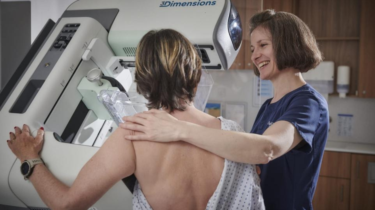
OneWelbeck mammographer with patient

Is it safe to get your breasts screened after getting implants? 

It’s a frequently asked question that is often met with a lot of misinformation.

“Women with augmented breasts deserve to be educated regarding their body and how to look after themselves, and this includes regular preventative screening for breast cancer,” says Professor Zoe Winters, a consultant breast surgeon and breast specialist from in London.

Below, Professor Winters addresses common concerns and debunks some myths about breast screening for women with implants.

Q: Can I get my breasts screened if I have implants?

A: With the misinformation about added risks and side-effects surrounding breast implants and screening, it’s understandable why women may feel anxious about getting checked. However, I would like to clarify that there are no added risks to the implants, and screening is a crucial step in detecting cancer. Implants consist of high-quality, cohesive silicone which is not susceptible to rupture under compression, and so the perception that they are at risk during a mammogram is false.

Radiologist ultrasounding the axilla

It is, however, true that there is a slightly different screening process. At OneWelbeck, we can discuss the process in detail with each patient and tailor the methods for screening to their age and implant location, where an implant is placed either on top or below the pectoral or chest wall muscle. If a patient has implants and is taking Hormone Replacement Therapy (HRT) for menopausal symptoms, we can also safely accommodate for this.

Q: How can I get my breasts screened safely with implants?

A: Screening a patient with breast implants requires an experienced mammographer who knows how to position the implant in the appropriate way to achieve an accurate image of the breast tissue, especially relating to the back part of the breast.

It is important to be aware that it’s often not possible to screen 100 per cent of the breast, but the more experienced a practitioner is, the better chance there is of getting a more accurate and representative image.

At OneWelbeck, we have optimised the way that we screen women with augmented breasts. The routine two views are performed giving a cross sectional view of the breast tissue followed by a method called the Eklund Technique, which is specifically designed to assess more than 90 per cent of breast tissues in relation to implants.

Q: Am I at higher risk of getting cancer if I have breast implants?

A: No, not at all. Risk factors can be determined by several factors, but augmentation surgery will not increase a woman’s likelihood of getting breast cancer. However, it is important to emphasise that women with implants are not protected from breast cancer. Cancer can occur anywhere within the breast tissues, including right at the back of the breast, towards the chest wall muscle. These cancers may be missed if the described imaging techniques are not applied.

Mammographer reviewing breast images

Lesions and lumps in certain areas may go undetected, which is why it’s so important to be screened by a mammographer with a high level of expertise. Issues with the implant itself, including implant rupture, also called a gel bleed, resulting in free silicone spreading across the breast, can also complicate imaging, running the risk of missing something.

The specialists at OneWelbeck will assess the outer silicone layers of the implants for disruption, internal silicone fractures and gel bleeds. Imaging will also include the armpits.

Q: Do I need to get my breasts screened any more or less frequently with implants?

A: No, the advice remains the same. The European guidelines recommend that women undergo breast screening annually from 40 years of age, and two yearly from 50 years onwards, regardless of implants. Currently, the UK NHS screening programme commences mammography from 50 to 74 years at three yearly intervals.

Receptionist at OneWelbeck

Women who routinely get screened (every one or two years) will have a significantly reduced chance of breast cancer related mortality, as we can often detect early cancers that cannot be felt during clinical examinations.

Q: What breast screening options are available to women who have had breast surgery?

A: At OneWelbeck, we will begin with a mammogram, which is an X-Ray of the whole breast that enables the detection of any lumps, anomalies or concerning issues. Every woman will have their breast tissue density score provided in their imaging report. Women need to be aware that their breast density can influence their risk of getting breast cancer, with the densest breast tissues increasing risks compared to those ladies with fatty breast tissue.

If a woman’s breast tissue is too dense (with or without implants) to be accurately screened with a mammogram, then we would recommend a breast MRI and bilateral breast ultrasound, which are specifically designed to identify abnormal lesions.

Breast surgery can increase scar tissue formation, causing the death of breast fatty tissues, and so the ability to compare current versus previous mammograms is important in order to assess changes over time.

The Resident in partnership with OneWelbeck

Talk on HRT and Breast Cancer with Kate Panter

Panel discussion on Breast Cancer, Menopause and HRT

Leading Clinicians Collaborate in an In-depth Panel Discussion on the link between Breast Cancer, Menopause and HRT

As we close Breast Cancer Awareness Month (October 22) a group of leading clinicians took part in a panel to discuss Breast Cancer, Menopause and HRT, (Hormone Replacement Therapy) tackling some of the myths associated with the subject whilst championing the emergence of a pioneering screening process and existing innovative diagnostic procedures available to patients offering greater precision in the treatment of breast cancer. (Click here to watch) or see below under media.

Between 1 in 7 women in the western world will get breast cancer in their lifetime. Many women now understand that if they are asymptomatic, but carry a BRCA mutation they are at a high lifetime risk of developing cancer.

Professor Zoe Winters, Senior London Consultant Breast Cancer Surgeon at New Victoria Hospital contributed to the discussion by sharing news that HRT produces huge overall health benefits that particularly derive from Oestrogen replacement at the time of perimenopausal symptoms. Safest forms of Oestrogen comprise transdermal administrations that don’t significantly raise risks of breast cancer in the long-term. It is Progesterone long-term that does this, where it is required in women with an intact uterus to reduce unopposed oestrogen effects on the uterine lining.

All women should be aware that we are not all at the same risks for breast cancer within the general population. We are currently guided by predicted averages in the general population. Based on a recent UK randomised trial, including European guidelines, we recommend commencing mammograms at 40 years of age annually. Fifty-year olds may safely undergo 2 yearly mammograms indefinitely.

A mammogram is performed to assess both breasts in total and by comparing it to previous mammograms. One key finding to communicate with each woman is “What is your breast tissue density?” This is quantified by the radiologist assessing the ratio of breast duct tissues that appear “white” compared to fatty tissues that appear “darker” on the mammogram. Breast tissue density is a strong predictor of one’s breast cancer risks, where very dense breast tissues raise your risks by 4-6-fold compared to very fatty breasts. Very dense breast tissues may also reduce the sensitivity of mammograms, and we may miss detecting 20% of breast cancers. However, this guides how frequently we recommend mammograms, and whether we include bilateral breast ultrasounds or breast magnetic resonance imaging or MRIs.

Dr Kate Panter, Obstetrician and Gynaecologist at New Victoria Hospital, Consultant Gynaecologist and a specialist at the British Menopause Society went on to discuss the taboos around both menopause and taking HRT. While she feels the peri-menopause itself can still be considered taboo since many women struggle to even admit they are experiencing symptoms, she explains: “The taboo around taking HRT has in large part, come from the way the media talks about breast cancer risks associated with HRT. They portray HRT as the only risk for breast cancer, and a huge risk at that, when in fact there are other more significant lifestyle factors, such as obesity for example that can put you at risk of breast cancer. “

Professor Barbara McGowan, an Endocrinologist agreed by saying that many women have been too scared to take HRT because the link between HRT and breast cancer has been greatly distorted by the media over the past 20 years. She went on to explain: “There are recent publications that suggest a slight increased risk of breast cancer with taking HRT but that it will depend on the type of preparation. Never- the- less that risk remains reasonably low, and, after stopping HRT that risk tends to regress in time”.

There is often confusion from a woman’s perspective about the various types of HRT available and knowing what would suit them best.

Dr Panter talked about the different administrations of HRT. She explained: “When a woman is peri-menopausal, or going through the menopause, within a year of reaching menopause, or immediately post-menopausal, the time when you are no longer menstruating, you need to take cyclical HRT.

This means taking Oestrogen throughout the month and progesterone for the second half of the month. If your periods have stopped, for approximately a year – then we can give you what we call continuous combined or no-bleed HRT, where we give you a little bit of oestrogen and a little progesterone every day.”

There are various ways to take HRT, options include taking HRT orally or transdermally (through the skin, like gel or patches) or a combination, oestrogen gel applied to the skin and progesterone tablets.

Dr Panter discussed the benefits of taking transdermal HRT: “HRT used to be given orally, but that’s no longer the case, now we prefer to give oestrogen through the skin, as it doesn’t have to withstand digestion and so we can give an exact copy of your hormone, a bio-identical form of HRT, one that more closely represents our body’s own hormones, so there are benefits to transdermal administration. A natural copy of progesterone, Utrogestan, can be taken orally. There are also synthetic progestogens to be taken orally and they are much stronger than the hormones our bodies produce.”

There are alternatives if women decide they don’t want to use HRT or can’t take HRT for example if they have had hormonal cancers, these include vaginal lubricants and non-hormonal vaginal moisturisers as the vaginal area can be dry during menopause and Selective Serotonin Reuptake Inhibitors (SSRIs) anti- depression drugs which are effective for night sweats and CBT or Cognitive Behavioural Therapy for anxiety.

Dr Panter explained: “By the time most women come to someone like me they have already tried to manage their symptoms, they’ve visited the health shop, tried Magnesium, Red Clover, spent a lot of time and effort on diet and exercise and are very often at the end of their tether. One of the things I do recommend, is a self-help guide to Managing Hot Flushes and Night Sweats: A cognitive behavioural self-help guide to the menopause written by Melanie Smith and Myra Hunter a professor from King’s College London, which I think is helpful for women to work through as it helps to think about our symptoms differently and CBT is incredibly effective at managing depression and anxiety. “

Professor Winters spoke about symptoms that women should be aware of before visiting a one-stop-clinic such as New Victoria Hospital for a triple assessment to determine if they have breast cancer:

A discrete or a perceived lump in the breast, that can be differentiated from the surrounding firm but normal dense breast tissues, and the intervening soft fatty tissues. The one-stop breast clinic assessment requires careful clinical examinations and explanations alongside complimentary breast imaging interpreted by expert consultant radiologists. She explained that over 95.5% of breast pain is benign and rarely related to breast cancer.

Other key symptoms to be aware of are any bloody nipple discharge or ongoing nipple discharge that could also be exacerbated by HRT, however, any sustained inflammatory changes to the breast skin or nipple skin should be investigated.

Professor Winters explained: “In a one-stop setting, we are teaching women how to examine their breasts and trying to be very clear about what a lump constitutes, compared to what normal breast tissues feel like.”

On the topic of how long to take HRT and when or of to stop taking it, Dr Panter said if you are going to take HRT the maximum benefit and the fewest risks are to start it early, so around the time of your menopause to derive the most benefit and least risk, particularly if you start it early, where there is no benefit on holding out.”

Dr McGowan reiterated during the session that the benefits far outweigh the risks and urged women not to be scared to use HRT if it’s appropriate in their case. Dr Panter explained that HRT will help our lifestyles and more holistically will help us to sleep, to be calmer and to cope at work and at home. Professor Winters said she looked forward to a more personalised and finessed breast screening processes as breast cancer care evolves in the future.

Biographies: Contributing Guests

Professor Zoe Winters – Breast  Cancer Consultant Surgeon & Breast Specialist

Professor Barbara McGowan – Endocrinologist

Dr Kate Panter – Consultant Gynaecologist at the New Victoria Hospital

With thanks to New Victoria Hospital and JFPR Consulting for putting this together.

Images of dense breast

Breast Density Updated 24/04/2026- By Prof Zoe Winters

Dense breasts and what this means for women undergoing breast screening

Updates on breast screening in women with dense breast tissues 24/04/2026

Three different supplemental imaging techniques have been compared in a UK randomised clinical trial called the BRAID UK trial https://www.thelancet.com/journals/lancet/article/PIIS0140-6736(25)00582-3/fulltext

The level 1 or highest quality evidence shows that when compared to standard mammograms combined with ultrasound:

  1. Contrast-enhanced mammogram (CEM)
    Or
  2. Abbreviated breast MRI

Are: 

Equally effective in detecting new breast cancers

Detect 3 times as many invasive cancers 

Detect earlier stage cancers half the size; and may influence / improve survival outcomes

Frequently asked questions:

1. Does having dense breasts increase my risk of breast cancer?
  • Increased breast density (C or D) increases risks of developing breast cancer compared with predominantly fatty breasts
  • Increased risk is moderate and requires consideration alongside other risk factors: family history, prior biopsies, and genetic predisposition
2. Should all women with dense breasts have supplemental screening?
  • Supplemental imaging should be discussed as part of shared decision-making
3. What are the main supplemental imaging options?
  • Contrast-enhanced breast MRI (Full MRI or “abbreviated/fast” MRI)

  • Abbreviated MRI takes 15 minutes and is equally effective and less costly

  • Contrast-enhanced mammography

  • Automated breast ultrasound aided by a machine rather than hand-held is less effective compared to MRI or contrast mammogram
4. How do I know if I personally should have supplemental imaging?
  • Shared-decisions are best guided by your 5-year risks of developing breast cancer described as High risk (greater than 1.66% and less than 6%) or Very High risk (greater than and equal to 6% at 5 years) 

  • You and your Consultant Breast Cancer Surgeon should weigh potential benefits (earlier detection) against harms (false positives which means “over detection” where a lesion that enhances is not a cancer; including potential increases in anxiety; and in costs to clients and providers) 

What is a Contrast-Enhanced Mammogram or CEM?

This is a mammogram done after injecting an iodine-based contrast dye into a vein, so that the scan can highlight areas with increased blood flow, such as many cancers 

It uses the same X-ray exam as a standard mammogram, but takes paired low energy and high energy images and then combines them to create an iodine-only image that shows enhancing areas more clearly.

CEM can help detect cancers that are harder to see on a standard mammogram, especially in dense breasts (C or D). It can also be used in circumstances to assess the extent or true size of a known cancer, to check symptoms or to assess the response to upfront medical treatments like chemotherapy or endocrine treatments like Letrozole. 

Practical point:

It is not the same as a 3-D mammogram which is called Tomosynthesis. It is the contrast that adds the extra information.

How does it compare with breast MRI?

Neither is universally “better”

Diagnostic performance:

CEM has a higher specificity (77%) which means fewer recalls and false positive biopsies than MRI

CEM has a lower sensitivity and cancer detection of 61% versus 100% for MRI

SO there is a slightly better overall discrimination for MRI (higher diagnostic odds ratio)

MRI is still preferred for: 

  • High-risk screening (High risk genes like BRCA; TP53; PALB2)

  • Detailed local staging like:

  • Multifocal / multicentric cancers 

  • Contralateral occult cancer which means cancers not seen on mammogram, like invasive lobular cancers (ILC) or non-calcified DCIS or ductal cancer in situ which is defined as intraductal or microscopic pre-invasive breast cancer cells 

  • Implants and lesions close to chest wall / axillary tail (upper outer breast tissue that extends into the axilla / armpit)

Practical points and patient-centred factors:

  • CEM is cheaper, quicker and easier to implement in mammography units where MRI may not be readily accessible

  • Women report a preference for CEM as there is no tunnel, and they feel less claustrophobic

Safety and contraindications:

  • CEM uses iodinated contrast plus iodinated radiation 

  • It is not advised where there is a significant iodine allergy; untreated overactive thyroid called hyperthyroidism; requires caution where there is renal / kidney impairment 

  • It is generally avoided for high-risk mutation carriers as a primary screening tool because of radiation

    *End of update*

June 2022

General importance

The density of breast tissues relates to the amounts or relative proportions of normal breast duct tissues that appear as the white component on an X-Ray or mammogram. This breast duct tissue is referred to as fibroglandular that means breast ducts surrounded by fibrous or connective tissues (collagen, fibrinogen). Connective tissues give structure and support to the breast ducts. It is the fibroglandular tissues that are assessed for any abnormal changes on mammograms.

Fibroglandular tissues absorb X-Rays and project as “white” on the mammogram. This is referred to as “dense” tissue. The amounts of fibroglandular tissue are compared to the amounts of fatty tissue that projects as “dark” on the mammogram as fat cells are mostly liquid, and are not dense like fibroglandular tissues. The amounts of fibroglandular tissue are largely genetically determined, and also depend on hormonal stimulations such as the oral contraceptive pill, the intrauterine Mirena coil and hormone replacement treatment (HRT).

Most cancers absorb X-rays to a similar extent as fibroglandular tissues, and therefore they also appear as white “masses” on a mammogram. Dense “white” tissues can therefore hide similar dense “white” cancers. (It’s like looking for a white ball in a snowstorm). This means that mammographically dense breasts have a reduced sensitivity (reduced detection of breast cancers) when relying on a mammogram only to detect breast cancers. In these cases, it is crucial to recommend additional screening using bilateral breast ultrasounds or breast MRI (Magnetic Resonance Imaging).

Assessments

A mammogram is the international standard for breast screening by “taking a picture” of each breast as a whole. The mammogram measures the amounts of fibroglandular tissues. The principle of an X-ray is to identify the “white” from the “dark” areas to calculate the ratio of how much “white” to “dark” areas there are in both breasts.

It is not possible to assess overall breast density using breast ultrasounds. The latter is operator-dependent and doesn’t take a picture of the whole breast, and is used to focus on a particular lesion seen on the mammogram. Ultrasound can’t report overall fibroglandular density in the breasts.

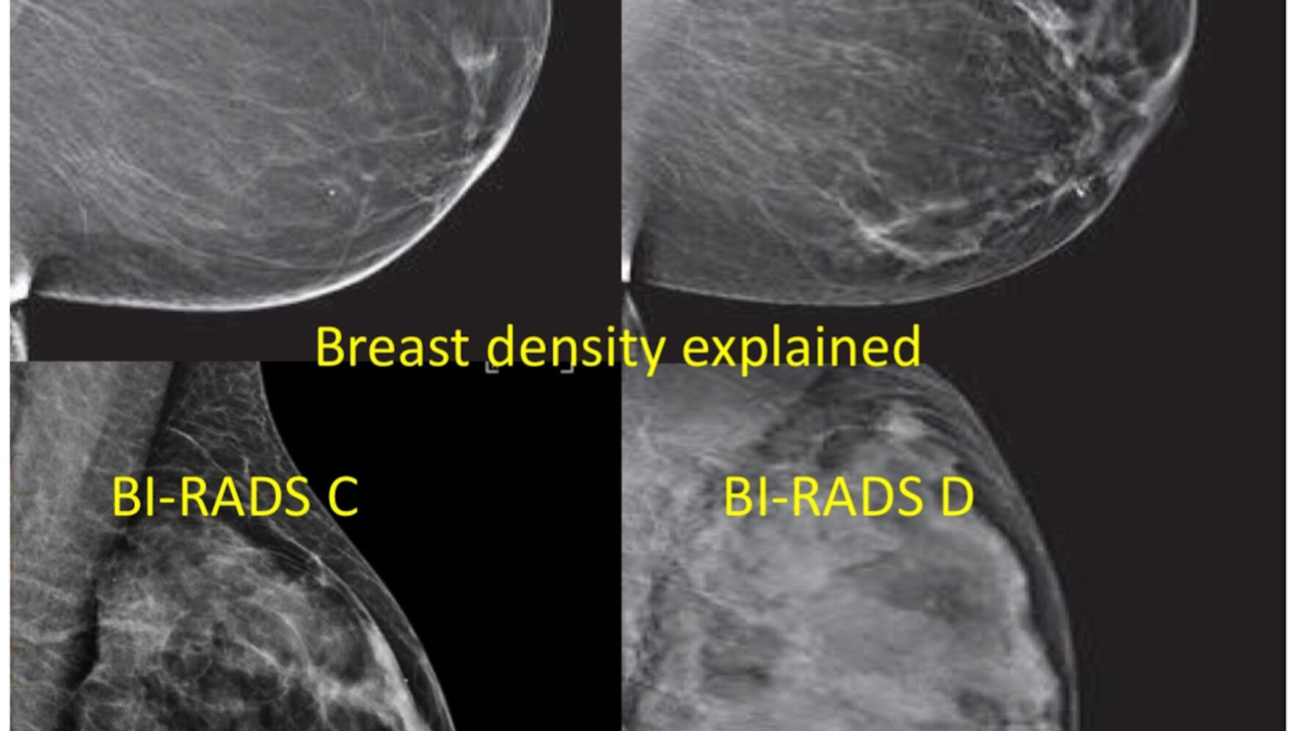
Below is a picture that shows how a cancer would present on a mammogram in each of the breast density categories.

In a fatty breast (A or B) a small cancer is easily seen, compared to a dense breast (C or D), whereby a large cancer is difficult to see.

Definition and measurements

Mammographic breast tissue density is classified into 4 categories by the ACR American College of Radiology and BI-RADS Breast Imaging – Reporting and Data Systems as follows:

Breast tissue density is reported from A to D by the Consultant Radiologist as follows:

A

The breasts are almost entirely fatty

about 10% of the screening population

B

Scattered areas of fibroglandular density

about 42% of the screening population

C

heterogeneously dense, that may obscure small masses

about 40% of the screening population

D

extremely dense, that lowers the sensitivity of mammography

about 8% of the screening population

Picture of category A, B, C, or D of breast density

ACR BIRADS C and D are clinically regarded as dense breasts

Radiologists measure the breast density on mammograms using different methods. Most commonly the Radiologist “eye-balls” the films and visually quantitates proportionate areas of “white” compared to “dark” tissues. Alternative methods use different automated computer programs potentially reducing variabilities in the reporting by Radiologists.

Implications

Dense breasts are a strong independent risk factor in all women and are currently used for breast cancer risk prediction. Dense breasts (ACR-C and D) raise breast cancer risks two fold above the current risks of 1 in 7- 8 women developing breast
cancer. This rate equals that of having a first degree relative such as either a mother or sister having breast cancer. Dense breasts account for 26% of cancers in post-menopausal women.

Extremely dense breasts (ACR-D) raise breast cancer risks by 4 to 6 fold compared to extremely fatty breasts (ACR-A).

A personal breast cancer risk test called MammoRisk uses an Artificial Intelligence (AI) – developed algorithm to assess breast cancer risks at 5 years. Breast density is one of the strongest prognostic factors for increasing breast cancer risks that is integrated into the MammoRisk test.

Recommendations by the European Society of Breast Imaging (EUSOBI)

X-ray based imaging techniques are all significantly affected by dense breast tissues leading to an under-diagnosis of breast cancers. The diagnostic sensitivity (optimal breast cancer detections) of mammograms is highest in women with fatty breasts (ACR-A) at 86- 89%, compared to women with dense breasts where the detection of breast cancers falls to 62-68%.

Current European recommendations are to perform bilateral synchronous ultrasounds with the potential to increase the overall imaging sensitivity from 77% to 91% in women aged from 40-49 years. Overall, however, ultrasound may be limited compared to performing contrast enhanced breast MRI. Supplemental MRI in addition to mammograms detected an additional 16.5 cancers per 1000 screening episodes.

Breast MRI assesses increases in tissue blood flow where cancers that comprise faster growing cells “light up” significantly more than normal or benign tissues like cysts or fibroadenomas. Results of a Dutch clinical trial called the DENSE trial ultimately concluded that a potentially more cost effective screening strategy in women with dense breasts would be to recommend 2 yearly mammograms and MRI that saves 8.6 lives per 1000 screened women. This is an evolving field and “we should watch this space”.

Sharing information with women

All women with dense breasts should have the advantages and disadvantages of enhanced breast screening using combined mammograms and MRI 2 yearly explained to them.

Advantages of enhanced breast screening:

Two yearly MRI leads to early cancer detection in about 10% of women and reduces the risk of dying from breast cancer to a little over 3%, providing a mortality reduction by about 40 %. A woman gains on average 15 years in good health, before she dies of another cause.

Disadvantages of enhanced breast screening:

Enhanced screening with breast MRI in particular will increase the chance that she will at least once experience the situation of a ‘false alarm’, i.e. receive a positive screening test which, after appropriate assessment, turns out to be a harmless finding. Of all positive (abnormal) screening findings, only about 30% are really cancerous; this value is similar for mammography and for MRI.

All women should be made aware of the clinical implications and importance of each mammogram report classifying their individual breast tissue density score labelled from A to D. This is now a mandatory requirement to all screening mammogram reports.

Breast tissue density informs future screening recommendations such as bilateral breast ultrasounds and or breast MRIs at every screening round. This approach is preventative in women and will save lives.

Updated 25/09/2025

Click on the DenseBreast-info logo below for more medically-sourced resources that was developed by health care professionals for both providers and patients, to advance the education on the screening and risk implications of dense breast tissue.

Logo for dense breast organisation with link


                                                            …TIME TO EDUCATE THE WORLD…
                    CLICK THE PICTURE BELOW FOR GREAT RESOURCES ABOUT BREAST DENSITY

World dense breast day logo the last Wednesday in September with link to information

     World Dense Breast Day last Wednesday in September, sponsored by DenseBreast-info.co

Darling magazine article

Breast Screening and latest care – By Professor Zoe Winters

 Professor Zoe Winters – Talks About The Very Latest In Breast Cancer Screening And Care

Professor Zoe Winters is a Consultant Breast Cancer Surgeon and Breast Specialist
I am delighted and honoured to share my passion and short story and how I got to where I am now in my career. My early passions in medical school related to acquiring as much knowledge as I could where I felt that “ knowledge is power” and can embolden how we present complex cases of breast cancer in the context of a fascinating expansion in the field of understanding different types of breast cancers, what causes breast cancer and how they might have arisen.

Professor Winters in theatre with female colleagues

In my current full time private practice in London, we talk a lot about gene mutations that occur at the time of the “egg and sperm” recombination to produce what are known as germline mutations. These account for the minority of breast cancer (8-10%), however the ability to test extended gene panels beyond BRCA1 and BRCA2 have changed our abilities to perform young onset breast cancer screening using tests like breast MRI including 3D mammograms and ultrasounds. These include genes like PALB2, CHEK2 and ATM whereby young age commencement of breast screening from 30 years using breast MRI can reduce breast cancer deaths by 56% to 60%.

LET’S LOOK AT THE GENES

On the other hand, we are passionate about talking about how spontaneous breast cancers occur through what we refer to as multiple functional changes in background genes that effectively act like the “body machinery”. We can assess multiple functional changes in 76 or more low risk genes to assess the changes in a “big gene dataset” using a validated personal breast cancer risk test called MammoRisk. This can allow us to predict personal breast cancer risks at 5 years and allows us to recommend the frequency of screening mammograms in all women from 40 years of age and older. This means we can personalise how often we recommend each individual woman’s mammograms with or without synchronous breast ultrasounds depending on their mammogram assessments of breast tissue density.

My passion is trying to deliver “state of the art knowledge” to my patients in a One-Stop rapid diagnosis breast clinic on all aspects of breast health and breast cancer symptoms that allows each individual to understand basic mechanisms that help to explain what we do and why.

MEDICINE IS MY PASSION

“Medicine is my absolute passion and I try to share my excitement and knowledge in each patient interaction”.

“I want women to feel empowered and comfortably reassured that we are working in a multidisciplinary team of experts ranging from Radiologists, to Pathologists and Breast Surgery that also encompasses more complex plastic surgery breast reconstructions and breast reductions and is always supported by clarifying surgery options and explaining the pros and cons of each type of surgery”.

“We share every medical report with each patient and place the patient at the centre of each decision-making step”.

“Breast Surgery has changed dramatically and is now synonymous with targeted personalised approaches based on each patient’s particular breast cancer biology. The aims of modern breast cancer surgery are to de-escalate the extent of surgery by combining the latest medical treatments such as chemotherapy and endocrine treatments (drugs that block the oestrogen and progesterone receptors in breast cancer) only when they are needed based on the cancer genes that can be measured in each cancer”. The patient’s age and cancer biology can also determine whether we give post surgery radiotherapy and how much we give”.

“We offer the latest techniques in localising small cancers using a Magnetic Seed, the size of a grain of rice”. The cancer is then detected using a Magnetic probe during surgery. We perform localisation of the axillary predictor lymph nodes using a liquid containing magnetic iron filings called Magtrace, that also allows us to perform diagnostic excisions of small numbers of sentinel lymph nodes”. The latter represent the key predominant draining lymph nodes that we would like to test diagnostically and determine whether we treat the axilla using axillary lymph node dissection that causes most of the side effects or whether we can treat the axilla using radiotherapy that causes half the numbers of side effects.

“We combine targeted cancer surgery with the latest breast remodelling techniques that are called” Therapeutic Mammoplasty” or “Therapeutic Breast Reduction” that are techniques that allow us to remove one or multiple cancers and optimally save the breast taking cognisance of the overall cosmetic results”.

SHE’S STILL ONE OF THE VERY FEW WOMEN LEADING IN THIS FIELD

“One of the highlights of my career was becoming a Professor of Breast Surgery which took 17 years and unfortunately numbers very few women in the UK. This pinnacle was the culmination of decades of work and dedication, but also pure “sweat and tears” in the context of a climate of patriarchal attitudes and discrimination at all levels of clinical and academic medicine”.

“I have used all of my experiences to power on because no one can snuff out passion and determination, and today I am thrilled to be part of the London Clinical fraternity of breast cancer surgery and breast health”. I truly work with some of the leading and top colleagues in London and Surrey”.

“One of my other passions is mentoring the future generation of surgeons so they can attain their full potential based on each individual’s talents. It was a privilege and honour to be able to do postgraduate research in molecular biology at Oxford University. I found it fascinating and that it underpins major advances in past and future drug developments and in garnering some understanding into mechanisms of breast cancer development and growth”.

BIOGRAPHY:

Professor Zoe Winters became a Professor of Breast Surgery in 2017 through University College London. She is an experienced, Senior London Consultant Breast Cancer Surgeon and Breast Specialist, specialising in the diagnosis, treatment and management of breast diseases, breast cancer and all benign breast problems. She is a nationally and internationally recognised senior breast surgeon, with clinical excellence awards through iWantGreatCare in 2018, 2019, 2021 and 2022. Zoe also received the London Prestige Health award for Top Breast Surgeon of the year in 2021 and 2022. Professor Winters trained in Johannesburg and Cape Town (South Africa) and completed her Doctor of Philosophy degree at the University of Oxford by receiving the prestigious Oxford Nuffield Senior Research Fellowship.

Professor Winters works closely with Oncologists, Geneticists, Plastic Surgeons and Psychiatrists at the Royal Marsden NHS Foundation Trust. Zoe has received many awards for her clinical research, including the King James IV Professorship for surgery research from the Royal College of Surgeons of Edinburgh. She has also led two world first randomised trials in breast surgery and has validated the first European Patient Reported Quality of Life questionnaire in breast reconstruction after cancer surgery that has been used internationally. Zoe now teaches post-graduate surgeons in the latest aspects of breast cancer and breast cancer surgery, including breast reconstructions.

You can find out more at the following sites:

Darling Magazine UK

New Victoria Private Hospital

HCA – Chelsea Outpatient Centre

OneWelbeck Women’s Health

iWantGreatcare
Doctify

You may also enjoy reading Michelle’s breast cancer journey under Professor Winters’ care

Patient story

The Story Of One Woman’s Breast Cancer Journey

When the COVID-19 pandemic kicked off in 2020, no one knew how far reaching the repercussions would be. Now, over a year in, we are beginning to understand. Macmillan estimates that due to the huge strain put on hospitals across the UK, there could now be up to 50,000 missing cancer diagnoses in the UK but, as anyone who has been impacted by cancer will know, the people tackling the disease are more than just a number. This Breast Cancer Awareness Month in October, we follow one woman’s journey with breast cancer from diagnosis to recovery with the help of private, charity-run medical facility New Victoria Hospital.

DISCOVERY

Receiving a cancer diagnosis in the middle of a pandemic lockdown with a teen child in need of home-schooling may sound like a nightmare, but this is exactly what happened to personal assistant Michelle Tatarczuk, 42, who found a lump in her right breast back in May 2020.

Obviously worried, she was keen to get it checked as soon as possible, but due to the COVID restrictions that were in place at the time she had to wait for a mammogram. Michelle adds, “I felt slightly concerned about the lump, but thought it would be ok as the GP said it felt benign and also because I have no history of breast cancer in my family.”

It wasn’t until October that Michelle was able to attend an annual health assessment. From there, she was quickly referred to Professor of Breast Surgery, Zoe Winters.

DIAGNOSIS

A nationally and internationally recognised breast surgeon, Professor Winters is one of the top breast consultants at New Victoria Hospital and has won several clinical excellence awards. Also awarded for her clinical research, she has led two world-first randomised trials in breast surgery. As such, Michelle knew she was in safe hands.

Professor Zoe Winter says, “Surgery is both an art and a science where wisdom is required to consider the latest thinking and innovations in treatment inspired by these concepts to impact positively on women’s lives”

It was through a mammogram, ultrasound, biopsy and MRI pathway at New Victoria Hospital that she received her complete diagnosis – a Grade II Pleomorphic Invasive Lobular Cancer in the right breast.

Professor Winters adds, “clinically there were no obvious abnormalities, but mammogram and ultrasound detected an atypical lump measuring 6 mm that biopsy confirmed was a small cancer. Breast MRI showed further small nodules that allowed conserving surgery”

Michelle recalls, “Nothing was picked up on my mammogram, only during my ultrasound. I am forever thankful to the sonographer who saw the shadow and did the biopsies. It could easily have been missed. It was a big shock to find out I had breast cancer, as I don’t have a history of this type in my family,” Michelle continues “and attending the consultations was a bit overwhelming at first as there was so much information to take in.”

This, she says, was made easier by the expertise and care of Professor Winters, who explained the results to her scans and the best course of treatment in a way that was easy to understand. “She was always very patient, reassuring and positive and following our consultations and she sent me lots of information to read through in my own time, which was very helpful.”

For cancer patients – particularly those who have a long and difficult journey to recovery – working alongside a medical team that treats you as humanely as possible is a top priority. Thankfully, Michelle found this in Prof. Winters. ”I truly feel that she really cares about her patients and makes you feel that you are not just a number. I very much enjoyed my consultations with her. She really is an amazing surgeon.”

During this period, Michelle also found the added touches that New Victoria provided particularly helpful considering the ongoing situation with the pandemic. “The facilities were great, the environment was very welcoming and COVID regulations were followed. There was always parking available (free of charge) and I never had to wait too long to be seen for my scans and appointments.”

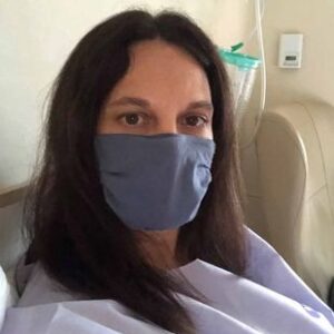
Michelle in Hospital recovering from surgery

TREATMENT

Michelle was recommended surgery, radiotherapy and injections as part of her treatment plan, with her operation taking place in December 2020 – two months after being diagnosed with breast cancer. Her Lumpectomy included a right wire guided local incision and dual localisation sentinel node removal.

Professor Winter says, “New localisation techniques allow the surgeon to remove very small cancers that can’t be felt. Likewise dye localisation of lymphatic draining lymph nodes allows simultaneous diagnosis and treatment of predictor lymph nodes called a sentinel lymph node biopsy. Early cancer detection predicts significantly improves clinical outcomes and ensures potential cures”.

Michelle was then given time to recover before starting on ten sessions of Radiotherapy.

RECOVERY

As anyone who has dealt with cancer knows, the road to recovery is rarely smooth sailing – particularly if it involves other challenging factors. For Michelle, the initial recovery from surgery took 8 weeks, which then had to be followed up with Radiotherapy, and a cocktail of daily and monthly injections – both of which she will need to continue for at least 5 years.

“The whole process – surgery and radiotherapy – was extremely difficult as I had to go to all consultations and treatment by myself and couldn’t see any of our family as we were in lockdown,” Michelle explains. “My husband spent a lot of time waiting for me in car parks!”

Speaking of the immediate surgery recovery, she said: “I was physically very sore and tired immediately following and was unable to do anything too physical for 8 weeks. I found this very difficult as I am a very active person.”

HEALING HURDLES

Cancer patients often find there are small hurdles to overcome on their road to healing – ones that are worth taking, but challenging nonetheless. Michelle experienced some muscle tightness in her arm and shoulder following her surgery and Radiotherapy, for which she underwent physiotherapy. She also had a slight skin lymphedema (a fluid build-up) and medication-induced menopause, causing hot flashes.

THANKFULNESS

Despite the huge challenge that Michelle’s breast cancer journey has presented for her, she is thankful for the time and health it’s afforded her. “These difficulties, as well as the lack of sleep they caused, did affect me physically and psychologically but are far better than the alternative.

“The treatment has removed all the cancer from my body. It saved my life.”
Early detection of breast cancer is vital as it increases the chance of successful treatment. For more information on the services provided by Professor Zoe Winters and New Victoria Hospital’s specialist breast clinic, head to the website here: New Victoria Hosptial.co.uk

Darling Magazine UK

Read Michelle’s story also in the Mirror

You may also like to read Darling Magazine Editor, Karine’s Cancer Journey

You may also like to read other cancer patient stories here

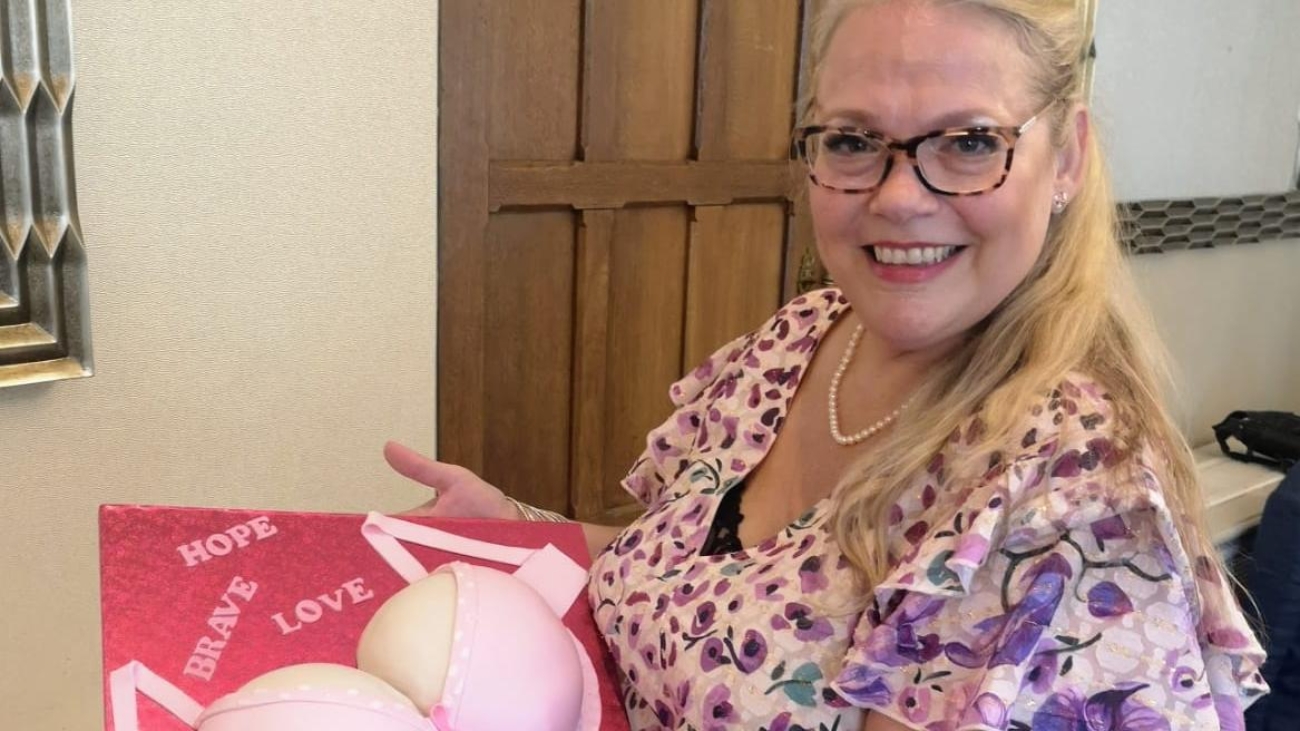
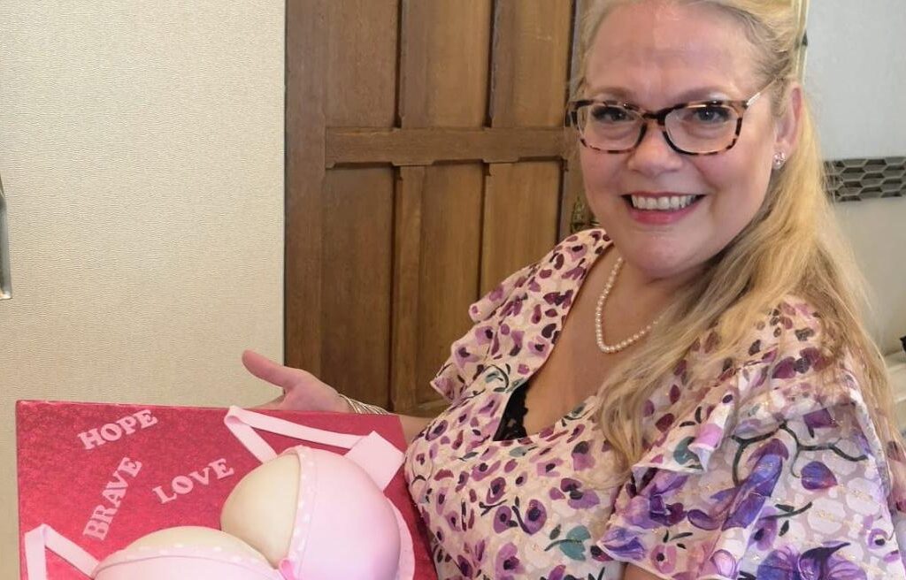
Patient bakes a cake for raffle

Breast cancer patient stories

Patient empowerment is essential both as a process and outcome. Breast patients, Mona and Andrea share their experience at New Victoria Hospital One-Stop Breast Clinic, highlighting the importance of making informed decisions.

Breast cancer affects 1 in 7 women in the UK. For Breast Cancer Awareness Month, we’ve spoken about the importance of cancer screening and prevention and the benefits of New Victoria Hospital One-Stop Breast Clinic in our previous blog article.

Today we would like to share with you the story of Mona and Andrea, two women who attended NVH One-Stop Breast Clinic and received very attentive care thanks to our multidisciplinary approach and fast track access to consultation and diagnosis.

Mona: the One-Stop Breast Clinic is my idea of less stress

“Before Covid, I was not aware of belonging to the 70+ group.

Unfortunately, I also was not aware, I should have had a yearly Mammogram test after the age of 70.

I detected the abnormality in my large breast by pure chance. After seeing my GP, I began my treatment under the NHS. I then decided to have a double Mastectomy at New Victoria Hospital under Prof Zoe Winters and her team.

It was a very successful experience. I like my new shape. The knowledgeable, trustworthy and caring treatment at NVH with its tasteful facilities made my recovery so easy.

I wish I had known about New Victoria’s One-Stop Breast Clinic earlier in order to have the choice to get the complete cancer treatment there.

Their Self Pay option is comfortable, and I didn’t have to worry about additional fees as it was an all-inclusive package.

Having the One-Stop Breast Clinic is my idea of less stress to resolve and treat the problem, more efficiently and quicker”.

Andrea: Maintaining control into a chaotic process

Andrea Knowles-Jones raising funds for Breast Cancer

“I have just turned 49 years old. I live in Surrey with my husband Andy and daughter Holly who will be 7 in November. I work for a large pharmaceutical wholesaler and am sole breadwinner, whilst Andy is a wonderful full-time parent doing the most important job of looking after Holly.

My business is extremely high pressured. I usually run at a million mph, and anything less than 100% just doesn’t cut it. Our colleagues are classed as key workers given that we deliver life-saving medication and essential healthcare products to the NHS, pharmacies and hospitals across the UK on a twice-daily basis and so when the Covid pandemic hit, we went into full crisis mode.

My annual screening mammogram came round in March.

I have yearly scans as my mother had breast cancer twice and then sadly passed away due to ovarian cancer. I was concerned when I was told I had pinky brown discharge coming out from my left nipple.

I was reassured that most likely nothing and indeed when I got the results a couple of weeks later, I got the standard ‘all OK, see you next year’ one-liner letter. I promptly pushed any worries to the back of my mind and continued focusing on work and the craziness that was ensuing in a Covid world.

A month or so later, however, I started noticing some pinky-brown spotting on bras and other clothing. I also found a small lump by my nipple. My breasts were feeling heavy and aching specifically on the left side, which was displaying symptoms.

Considering the recent clear mammogram result, I wasn’t overly panicky. I spoke to my GP who got me quickly referred back to the breast team who sent me for further tests.

The night before that appointment, I noticed a large hard area on the side of my breast, not a lump as such, but definitely noticeable. Not sure how I hadn’t picked up on it before. Again though nothing showed up on the mammogram, but the doctor was concerned about the hard area when she did the ultrasound and took biopsies there and then.

Two weeks later, I had a telephone consultation, and I was absolutely shocked to receive a diagnosis of DCIS intermediate level in my left breast. I honestly was not expecting it but went straight into pragmatic, action crisis mode. I pressed on with organising MRI and other tests so we could confirm the extent of disease and work out a treatment plan.

This was all done initially through an NHS consultant, but I am lucky to have medical insurance which when we got as far as we could with investigations being progressed on the NHS, I triggered.

As a result of the pandemic, local private hospitals were under the jurisdiction of the NHS, and so I had to look further afield to find a suitable consultant to look after me. This turned out to be a godsend and actually the silver lining of the Coronavirus situation as I was lucky enough to go under the care of Professor Winters.

KNOWLEDGE IS POWER

Obviously, trust is key, but you also need a level of chemistry with your consultant who you are going to be discussing and sharing some very intimate and often distressing situations with.

For me, knowledge is also power and I found the ‘being out of control’ aspect of my diagnosis incredibly challenging as in my personal and professional life I have to juggle a lot of responsibility and being organised, planned and in control is how I cope.

From my initial chat with Jenny, Prof Zoe’s practice manager to the moment I met Zoe in clinic at the New Victoria Hospital (the first clinician I had actually met face to face since my diagnosis), I knew I had made the right choice and was in the best hands.

Professor Winters was to me the perfect blend of providing informative and expert consultation taking the time to talk through all aspects of tests, scans, biopsies, results etc to ensure I understood everything. She showed empathy and understanding of what I was going through.

As a lover of detail myself, Zoe left no stone unturned and was beyond thorough to ensure we made informed decisions on my surgery and treatment plan. She gave me back my power and control and I will be forever grateful for that and cannot thank Professor Zoe enough.

I was so assured that whatever additional help and counsel was required, Professor Zoe will recruit and bring the very best experts on to the team to look after me. I really had and still have the best of the best on my side.

I still struggle with acceptance of everything that has been happening to me as at each appointment, things seem to worsen, and I had to take in more and more bad news to get my head around.

I have had a skin-sparing mastectomy of my left breast with immediate reconstruction and post pathology results revealed a large area of invasive cancer as well, which was a shock.

But we got it in time, and I am now doing all possible to prevent it coming back which has included radiotherapy and now hormone therapy which has required me to have medically induced menopause. I have more surgery ahead to help me ‘get my body’ back, but I have a top, first-class plastic surgery caring for me, thanks to Zoe.

I often think about what the situation could have been if I had just left things and not persisted with checks. What if I had left it because I was too busy and didn’t get round to it until next year’s annual check. Thankfully, I didn’t, and my clear message is that YOU know your body better than anyone else or any machine and so trust your gut instinct and if in doubt, check it out. It might be nothing, which is great but if it is something, you can get it early and do something about it.”

Andrea’s story in The Times