
A Consultant Radiologist will provide you and your Consultant Surgeon with an imaging report with numbered categories for each type of imaging.
At each stage of the One-Stop triple assessment, the suspicion for malignancy is graded to create an overall risk index, as described below. The key below is used to establish whether a lesion is likely to be a benign, indeterminate, or highly suspicious that requires further imaging and or an image guided core needle biopsy.
(P) = Physical / Clinical examination by the Surgeon (P1, P2, P3, P4)
(M) = Mammographic Grade Description (M1, M2, M3, M4)
(U) = Ultrasound Grade Description (U1, U2, U3, U4)
(B) = Biopsy Grade Description (B1, B2, B3, B4)
What the codes mean on the report:
P1 / M1 / U1 / B1 = Normal
P2 / M2 / U2 / B2 = Benign
P3 / M3 / U3 / B3 = Uncertain / probably benign
P4 / M4 / U4 / B4 = Suspicious of malignancy
P5 / M5 / U5 / B5 = Highly suspicious of malignancy
Quadrants: Right & Left
Each breast is divided into 4 quadrants as the surgeon faces the patient with the nipple areola in the centre. The right breast correlates with the client’s right, and the left breast with the client’s left. The Radiologist and Surgeon each describe the quadrant of the breast where the lesion is seen, whether it is central or peripheral, and also include location details by referring to the numbers of the face of a clock as one is looking at it.
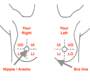
UO: Upper Outer (superior and lateral)
UI: Upper Inner (superior and medial)
LO: Lower Outer (inferior and lateral)
LI: Lower Inner (inferior and medial)
C: Nipple and Areola (Central with clock numbered position)
IMF: Infra-mammary fold or bra line (inferior central, inferior lateral, and inferior medial)
Terminology found in your imaging report(s):
Addendum = The supplementary text added at the end of a previously approved radiology report, to correct or expand on an original assessment by the Consultant Radiologist. Additional radiology review is required where there is a need to compare current images with previous mammogram or ultrasound images.
Architectural distortion = This suggests a suspicious or highly suspicious lesion on a mammogram that may require a tissue biopsy, but can also be caused by scar tissue after breast surgery. Occasionally, a benign lesion such as a radial scar comprises benign breast ducts (adenosis) with extensions like spicules associated with fibrosis (scarring) that is normal tissue in more than 90% of cases.
Asymmetric density = A mammogram lesion seen in one breast compared to the same location in the other breast, that may require further investigations, if it is new compared to previous imaging. This may also arise from differences in the amounts of breast duct tissues between the breasts that are part of developmental differences at birth, and usually requires additional imaging views using magnification mammogram and 3 D mammograms called tomosynthesis.
Axillae = Armpits
Benign = Non-cancerous
Bilateral = Both breasts
Calcifications = Evidence of coarse calcium deposits that develop in normal breast tissue such as cysts, and benign fibroadenomas, including blood vessels and breast duct. Calcifications may also occur in malignant lumps, if slow growing, or if fast growing following death of breast cancer cells called necrosis.
Complex breast cyst = Contains a solid tissue or polyp-like growth within the cyst wall, and requires a tissue biopsy.
Complicated breast cyst = This type of cyst has some semi-solid particles / fragments floating in the fluid on ultrasound, and requires either aspiration, or a core tissue biopsy if the aspirate is either “bloody”, or the cyst doesn’t disappear. A complicated cyst may also be called a glue cyst (see below).
A complicated cyst wall may also be thickened, and require a core tissue biopsy and not simply an aspiration.
Cortical thickness = The normal thickness for the lymph node cortex is 3 mm. If the lymph node cortex is thicker, it requires a tissue core biopsy to exclude abnormal cells. Often the thickened lymph node is a normal reactive node that represents a normal immune response after a biopsy or infection. However, it is important to exclude cancer cells.
Cyst = A cyst is a small cavity filled with fluid. Cysts are normal and arise from microscopic breast ducts as they age. Hormones like Oestrogen, Progesterone and Prolactin (milk producing hormone) can increase secretions by the breast duct cells that line the walls of the microscopic breast ducts. The breast duct walls become thinner with age causing the ducts to dilate called duct ectasia. The thin duct blows-out fluid filled cysts.
Cysts are mostly described as benign and are rarely associated with cancer. Most cysts resolve spontaneously, and are nothing to worry about. If the cyst is large or causing discomfort, your Radiologist may draw off the fluid using a fine needle and a syringe, called cyst aspiration.
Fibroadenoma = A benign (not cancerous) developmental lump that occurs mostly in puberty and is not a risk factor for breast cancer. Fibroadenomas do not become cancers. They have an unknown cause but comprise a mixture of fibrous and breast duct or glandular tissue. They may fluctuate in size with hormonal triggers like the pill, hormone replacement therapy, pregnancy and breastfeeding. One third increases in size, one third becomes smaller, and one third disappears. They do not require surgery removal; however, surgery excision may be offered as a choice if they increase in size to over 30 mm.
Fibrocystic changes = Normal breast duct tissues, admixed with fibrous connective tissues, and benign simple cysts.
Fibroglandular density = Normal overlapping breast duct tissues and fibrous tissue producing this appearance on the mammogram.
Findings = What was “found” from the radiology exam, listing each area of the breast that was examined in the diagnostic imaging study
Focal = This describes a change or finding in one area of the breast on imaging.
Glandular = Glandular tissue is another term for dense (increased breast duct tissue versus fatty tissue), normal breast duct tissue that appears white on mammogram and ultrasound. Greater amounts of glandular tissue occur in the upper outer breast areas, central nipple area, compared to the lower breast segments.
On clinical examination, glandular areas are firmer, compared to the fatty breast tissues that are softer.
Glue cysts = Contains semi-solid particles. It requires cyst aspiration to determine if it disappears completely in which case, it is a simple or normal and benign.
Hypoechoic mass = This means a lesion that is more dense or solid than usual as seen on an ultrasound scan. Ultrasound uses sound waves that are either absorbed (look black like a cyst which is fluid), or bounce off tissues (look white like a solid lump), based on their density. The sound waves form the black and white image you see on an ultrasound screen e.g. The whiter image is the breast duct tissue, and the darker image is fatty tissue or fluid that looks like a “black hole” in the case of simple cysts.
Indeterminate = Atypical
Indeterminate lymph node = The axillary lymph node may be larger compared to the other lymph nodes. The lymph node comprises a fatty centre surrounded by a rim of normal lymphatic cells called lymphocytes,that form a rim called a cortex. An indeterminate node may have a slightly thickened cortex that requires a lymph node biopsy if its thickness is greater than 3 mm.
Lipoma = This is a benign developmental growth of normal fat cells that forms a soft tissue benign lump with a defined capsule or border seen on ultrasound. They can be surgically removed if troublesome or increase in size, but lipomas in the breast do not increase breast cancer risks.
Large lipomas may require MRI soft tissue imaging to ensure that they are not malignant sarcomas called liposarcomas. The latter should be referred to a specialist Sarcoma clinic.
Lobulated = This refers to a benign fibroadenoma that has a smooth border with undulations called “lobules” on imaging.
Macrocalcifications = Appear as large white coarse spots that look like dashes or dots. They are rarely indicative of a cancer, and their presence does not require additional tissue biopsy testing or follow up, except routine breast screening.
Malignant = Cancerous
Microcalcifications = Appear as tiny finely clustered white specks. They are often referred to as appearing like “salt and pepper” on a mammogram. They are also usually non-cancerous, but if these tiny specks form certain patterns or have increased over time based on comparison mammograms, they can indicate a microscopic cancer that is confined to a microscopic duct called ductal cancer in situ (DCIS). Generally, further imaging, and evaluation is required using additional mammogram views and possible mammogram guided tissue biopsy.
Modified WHO criteria used = Form signed to say that allergies have been checked.
Nodular fibroglandular = This is described above under “Nodular Pattern”.
Nodular pattern = The normal breast is composed of dense fibrocystic breast duct tissues, that are intermixed with fatty tissues in a fairly irregular pattern that causes a nodular appearance on imaging, or a “lumpy” feel on clinical examination. Mostly, these are related to solid masses that appear white on mammograms and ultrasound in keeping with dense normal ductal breast tissue.
Fibrocystic changes that contain sub-centimetre cysts are diagnosed on ultrasound as multiple “small black holes” in keeping with cysts that are normal.
No focal solid masses = Normal breast tissues.
Palpable lump = A palpable lesion / lump in the breast is usually subjected to triple assessment (Clinical examination, Imaging and Core Biopsy or Fine Needle Aspiration).
Partially cystic mass = This means a semi-solid lesion that has mixed solid and fluid components. This may suggest a complex cyst that requires a tissue biopsy of the solid component if the cyst doesn’t collapse completely on fluid aspiration, of the cyst fluid is “bloody”.
Sarcoma = Rare form of rapidly growing soft tissue cancer that may arise after radiotherapy for breast cancer treatment, or is a malignant phyllode tumour, that is a variant fibroadenoma diagnosed on tissue core biopsy or following surgery excision.
Scattered microcysts = Less than 10mm in size. Too small to feel, and detected using ultrasound.
Scattered simple cysts = Cysts that can be felt are called symptomatic, and may be one or multiple in number. A breast cyst often feels like a mobile smooth grape, or a water-filled balloon, but sometimes feels hard. Breast cysts don’t require treatment, unless they are large and painful, which requires aspiration.
Sebaceous cyst = This is a benign developmental skin cyst that occurs in the dermis of the skin. It is not related to breast duct tissues. If troublesome, it requires a referral to a Consultant Dermatologist for skin ellipse excision under local anaesthetic.
Sub-centimetre cysts = Less than 10mm in size. These are not large enough to be seen on a mammogram, but are detected on ultrasound. They are benign and occur as part of duct ageing and breast duct secretions.
Subcutaneous (cystic lesion) = See sebaceous cyst.
Superficially = This means close to the skin and fatty tissues underlying the skin.
Tomosynthesis or 3-D Mammogram = Advanced form of mammography that uses a low-dose x-ray system and computer reconstruction to create three-dimensional images of the breasts. Tomosynthesis is used for further evaluation or very dense breasts, and is considered to be enhanced breast screening.
Unifocal = This means a single lesion.
Unilateral = Single breast
Unremarkable = If an area appears as normal breast tissue (there is nothing to worry about)
Vascular mass = This means lesion with increased blood flow seen on doppler ultrasound.
BI-RADS Mammogram Scoring – Understanding your BI-RADS Score
BI-RADS stands for Breast Imaging Reporting and Data System. It is an assessment tool used to rate the results of a mammogram test.
The BI-RADS score has seven levels of ranking:
- Category 0: Additional imaging is required to provide a category. The results were inconclusive.
- Category 1: Your test result is negative. There is no significant or noticeable abnormality on your mammogram. It is important to continue recommended routine screening.
- Category 2: A benign non-cancer lesion was identified. There is a benign calcification or fibroadenoma, but it is not of concern. A tissue core biopsy may be indicated.
- Category 3: A lesion was identified that is most likely benign. You may require a tissue core biopsy if the lesion is solid or a follow up mammogram at 6 months to monitor the area for changes.
- Category 4: There is a suspicious abnormality that may be cancerous. This result requires a tissue core biopsy.
- Category 5: The growth is highly likely to be malignant, meaning it is probably cancer. This result requires a tissue core biopsy.
- Category 6: A biopsy confirms the results of your mammogram, and the growth is identified as a cancer. Treatment should begin and be directed by a Consultant Breast Surgeon and Breast Specialist multi-disciplinary team.
BI-RADS reporting mammogram breast density
Your mammogram report will also include an assessment of your breast density, that describes the ratio between the amount of normal breast duct tissues and fibrous tissue called glandular tissues (whiter), compared to the fatty tissues (darker). The denser your breasts, the harder it can be to see abnormal areas on mammograms such as solid lesions, that can also appear white, making it difficult to separate a ‘white cancer” from normal dense breast duct tissues.
In a mammogram report, the radiologist uses a standard system called BI-RADS or ACR (American College of Radiology). The breast tissue density is classified into 4 categories from A-D. These categories describe the amounts of fatty tissues (seen as darker), compared to dense breast duct tissues (seen as whiter) in your breasts. The amount of breast tissue density increases from A to D, with D representing extreme breast tissue density as follows:
A: The breasts are almost entirely fatty with a greater proportion of darker tissues versus breast duct tissues that are whiter (about 10 % of the screening population).
B: The breast comprises mostly fatty tissue with a few scattered areas of dense fibrous and glandular breast duct tissues (about 42% of the screening population).
C: The breasts comprise multiple areas of fatty and dense breast duct tissues, that are referred to as heterogeneously dense, and may obscure small masses (about 40% of the screening population).
D: The breast is almost entirely comprised of dense breast duct tissues, and is referred to as extremely dense, that lowers the sensitivity of mammograms (about 8% of the screening population).